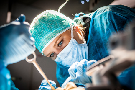
We develop and apply advanced photonics technology for surgical imaging, focusing on minimally invasive and robotic-assisted surgery.
What we do
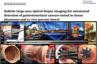
Our research focuses on developing and applying photonics technology for surgical imaging, including multispectral, polarization-resolved, and fluorescence imaging, and the use of fluorescently labelled gold nanorods for theranostics. We also work on illumination and vision systems for endoscopy combining miniature light sources (such as LEDs and laser diodes) with computer vision techniques for structured lighting and tissue surface reconstruction as well as the use of robotic guidance of optical probes.
Why it is important?
Better surgical guidance can improve the accuracy of surgery, reducing the time and cost to complete an operation as well as improving the outcomes for the patient.
How can it benefit patients?
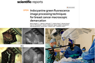
If the detection and diagnosis of cancer can take place live at the same point as the surgery is performed then in some cases there may a better chance for complete clearance without a need for further surgery. A good example of this is in our GLOW study, as described in this article and this podcast.
Summary of current research
- Fluorescence and diffuse reflection spectroscopy
- Multispectral imaging
- Multispectral structured lighting
- Near infrared fluorescence imaging
- Polarisation resolved endoscopy
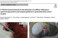
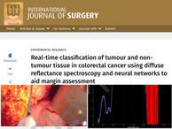
Fluorescence spectroscopy has been shown to be a promising approach for the analysis of diseased liver tissue. It is a fast, objective and non-destructive method to detect change in the endogenous fluorophores distribution and could prove to be a valuable tool for non-alcoholic fatty liver disease diagnosis and transplant graft assessment. Data has recently been acquired at the site of thermal tissue fusion with an RF anastomosis device by incorporating the probe into the device jaws.
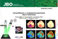
Multispectral imaging is involves collecting images of a target at different wavelengths to compile a spectrum for each pixel. We have developed endoscope and operating microscope systems that can be used in surgeries using standard clinical equipment and have translated these for in vivo use, including in humans. In surgical applications the device can detect ischaemia, although tissue motion artefacts need to be corrected using image alignment and deblurring algorithms developed with collaborators at UCL. We have worked with Mr Richard Smith’s team to monitor ischaemia during womb transplantation, and having recorded useful data during animal trials on sheep and rabbit models. We have also imaged many porcine small bowel samples during trials of a RF bowel anastomosis device, in collaboration with Covidien/Medtronic. We received ethics approval for imaging during human brain surgery in 2020.
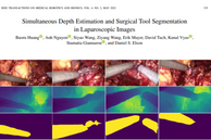
Three dimensional quantification of organ shape and structure during minimally-invasive surgery (MIS) could enhance precision by allowing the registration of multi-modal or pre-operative image data (US/MRI/CT) with the live optical image. We are developing flexible endoscopic multispectral structured illumination probes that label each projected point with a specific wavelength using a supercontinuum laser. Recent work has used spot shape for surface normal detection and calibration free operation, including in vivo, to make the devices more compatible with surgery.
Near infrared fluorescence can allow surgeons to target a particular tissue based on the accumulation of a contrast agent. Our home-built imaging system, called GLOW, consists of a camera device which uses illuminates a cancerous tumour during surgery and images the white light and near infrared fluorescence. This innovation will prevent people with breast cancer from having to endure another round of surgery. The adaptability of the camera system means that we can rapidly change the optical filtering to match different fluorophores and targets as they are developed. We are currently using the device in Breast Conserving Surgery with ICG and 5-ALA.
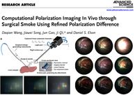
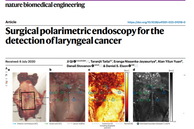
We have created a range of rigid endoscopic polarization (and wavelength) resolved imaging systems. These include simple cross-polarized imaging modalities, as well as full 3x3 and 4x4 Mueller matrix imaging, with either mono or stereo endoscopes. Creating the world’s first devices based on standard endoscopes has provided the possibility to rapidly translate the technology for imaging in vivo and ex vivo, the results of which suggest that the technique can reveal tissue microstructure and orientation, as well as enhancing the visibility of microvasculature. These aspects are being explored in the operating theatre, both on the back table and during imaging of peritoneal metastases.
Current research funding support
Recent papers
Selected recent papers. For a full list refer to my Imperial publications page, Orcid, or Google Scholar.
Current members
Professor Daniel Elson
/prod01/channel_3/media/images/people-list/180829-elson-daniel-005-tojpeg-1591262304362-x1_1609777271089_x4.jpg)
Professor Daniel Elson
Head of Group
Giulio Anichini
/prod01/channel_3/media/images/people-list/Giulio-Anichini.jpg.png)
Giulio Anichini
Clinical Research Fellow
Vadzim Chalau
/prod01/channel_3/media/images/people-list/Vadzim-Chalau.jpg)
Vadzim Chalau
Research Associate
Maria Leiloglou
/prod01/channel_3/media/images/people-list/Maria-Leiloglou.jpg)
Maria Leiloglou
Research Associate
Scarlet Nazarian
/prod01/channel_3/media/images/people-list/Scarlet-Nazarian.png)
Scarlet Nazarian
Clinical Research Fellow
Zepeng Hu
/prod01/channel_3/media/images/people-list/Zepeng-Hu.jpg)
Zepeng Hu
PhD student
Kaizhong Deng
/prod01/channel_3/media/images/people-list/Kaizhong-Deng.png)
Kaizhong Deng
PhD student
Naomi Laskar
/prod01/channel_3/media/imperial-college/data-science-institute/blank-profile-picture-973460_960_720.jpg)
Naomi Laskar
Clinical Research Fellow
Maxime Giot
/prod01/channel_3/media/images/people-list/Maxime-Giot-400X400.jpg)
Maxime Giot
Clinical Research Fellow
Naim Slim
/prod01/channel_3/media/images/people-list/Naim-Slim-400X400.jpg)
Naim Slim
Clinical Research Fellow
Nikita Chander
/prod01/channel_3/media/images/people-list/Nikita-Chander-400X400.jpg)
Nikita Chander
Clinical Research Fellow
Connor Daly
/prod01/channel_3/media/images/people-list/Connor-Daly-400X400.jpg)
Connor Daly
PhD Student
Alumni
Dr Kevin Koh (PhD graduate)
/prod01/channel_3/media/images/people-list/Kevin-Koh-400X400.jpg)
Dr Kevin Koh (PhD graduate)
Dr Tobias Wood (PhD graduate)
/prod01/channel_3/media/images/people-list/Tobias_Wood.jpg)
Dr Tobias Wood (PhD graduate)
Dr Vincent Sauvage (PhD graduate)
/prod01/channel_3/media/images/people-list/Vincent_Sauvage.jpg)
Dr Vincent Sauvage (PhD graduate)
Dr Lipei Song (PhD graduate)
/prod01/channel_3/media/images/people-list/Lipei_Song.jpg)
Dr Lipei Song (PhD graduate)
Dr Neil Clancy
/prod01/channel_3/media/images/people-list/Neil_Clancy.jpg)
Dr Neil Clancy
Dr Ji Qi (PhD graduate)
/prod01/channel_3/media/images/people-list/Ji_Qi.jpg)
Dr Ji Qi (PhD graduate)
Dr Rui Li (PhD graduate)
/prod01/channel_3/media/images/people-list/Rui-Li-(2).JPG)
Dr Rui Li (PhD graduate)
Dr Yi Cheng (PhD graduate)
/prod01/channel_3/media/images/people-list/Yi-Cheng.jpg)
Dr Yi Cheng (PhD graduate)
Dr Beatriz Garcia-Allende
/prod01/channel_3/media/images/people-list/Beatriz_Garcia-Allende.jpg)
Dr Beatriz Garcia-Allende
Dhurka Shanthakumar
/prod01/channel_3/media/images/people-list/Dhurka-Shanthakumar_300X400.jpg)
Dhurka Shanthakumar
Clinical Research Fellow
Dr Sinan Li (PhD graduate)
/prod01/channel_3/media/images/people-list/Sinan-Li.jpg)
Dr Sinan Li (PhD graduate)
Dr Clement Barriere
/prod01/channel_3/media/images/people-list/Clement_Barriere.jpg)
Dr Clement Barriere
Mr Shobhit Arya (PhD graduate)
/prod01/channel_3/media/images/people-list/Shobhit-Arya.jpg)
Mr Shobhit Arya (PhD graduate)
Dr Jianyu Lin (PhD graduate)
/prod01/channel_3/media/images/people-list/Jianyu-Lin-large.JPG)
Dr Jianyu Lin (PhD graduate)
Dr Lei Su
/prod01/channel_3/media/images/people-list/Lei-Su-large.png)
Dr Lei Su
Mr Taran Tatla (PhD graduate)
/prod01/channel_3/media/images/people-list/Taran-Tatla-portrait.jpg)
Mr Taran Tatla (PhD graduate)
Mr Mohan Singh (PhD graduate)
/prod01/channel_3/media/images/people-list/Mohan-Singh-2.jpg)
Mr Mohan Singh (PhD graduate)
Xueqing Sun
/prod01/channel_3/media/images/people-list/Xueqing-Sun.jpg)
Xueqing Sun
Dr Maria Elena Gallina
/prod01/channel_3/media/images/people-list/Maria-Gallina-400X400.jpg)
Dr Maria Elena Gallina
Dr David Harris-Birtill
/prod01/channel_3/media/images/people-list/David-Harris-Birtill.jpg)
Dr David Harris-Birtill
Dr Maria Tziraki
/prod01/channel_3/media/images/people-list/Mary_Tziraki_large.JPG)
Dr Maria Tziraki
Yu Zhou
/prod01/channel_3/media/images/people-list/Yu-Zhou.jpg)
Yu Zhou
Fernando Avila-Rencoret
/prod01/channel_3/media/images/people-list/Fernando-Avila.jpg)
Fernando Avila-Rencoret
Dr Mirek Janatka
/prod01/channel_3/media/images/people-list/Mirek-Janatka.jpg)
Dr Mirek Janatka
Dr Elham Nabavi
/prod01/channel_3/media/images/people-list/Elham-Nabavi.JPG)
Dr Elham Nabavi
Dr Ming Zhao (PhD graduate)
/prod01/channel_3/media/images/people-list/Ming-Zhao.jpg)
Dr Ming Zhao (PhD graduate)
Elizabeth Noble
/prod01/channel_3/media/images/people-list/Elizabeth-Noble.jpeg)
Elizabeth Noble
Martha Kedrzycki (PhD graduate)
/prod01/channel_3/media/images/people-list/Martha-Kedrzycki.jpg)
Martha Kedrzycki (PhD graduate)
Baoru Huang (PhD graduate)
/prod01/channel_3/media/images/people-list/Baoru-Huang.jpg)
Baoru Huang (PhD graduate)
Ioannis Gkouzionis (PhD graduate)
/prod01/channel_3/media/images/people-list/Gkouzionis,-Ioannis_300X400.jpg)
Ioannis Gkouzionis (PhD graduate)
Contact Us
The Hamlyn Centre
Bessemer Building
South Kensington Campus
Imperial College
London, SW7 2AZ
Map location
