Biosurgery and Surgical Technology performs research across a large portfolio of cross-cutting themes such as robotics, imaging, sensing, neuroergonomics, artificial intelligence, machine learning and computational biology. The surgical patient remains at the centre of our research philosophy through our global network of research collaborations.
Key research themes:
Themes within surgical technology
- Artificial intelligence and machine learning
- Endovascular robotics
- Frugal innovation and medical design
- Human-centred surgical technology
- Image-enhanced operating environment
- Intelligent surgical devices
- Neuroergonomics and perception
- Surgical imaging and biophotonics
- Surgical robotics
The development of artificial intelligence (AI) and machine learning (ML) techniques such as Artificial Neural Networks, Bayesian Networks and Decision Trees has begun to be incorporated into oncology to ‘learn’ from past examples and detect hard to discern patterns from complex data sets. Large amounts of information are now collected about patients and their cancers and AI programmes can analyse huge datasets to provide diagnostic support and to personalise treatments. For instance, AI methods could improve cancer diagnoses by using individual patient images in combination with digital libraries including proteomic and genomic assays. The UK has become a leader in AI research with London-based Google DeepMind considered to be the leading group of applied researchers in the world. DeepMind have built a close relationship with Imperial College and are looking at research opportunities across cancer pathways.
Researchers
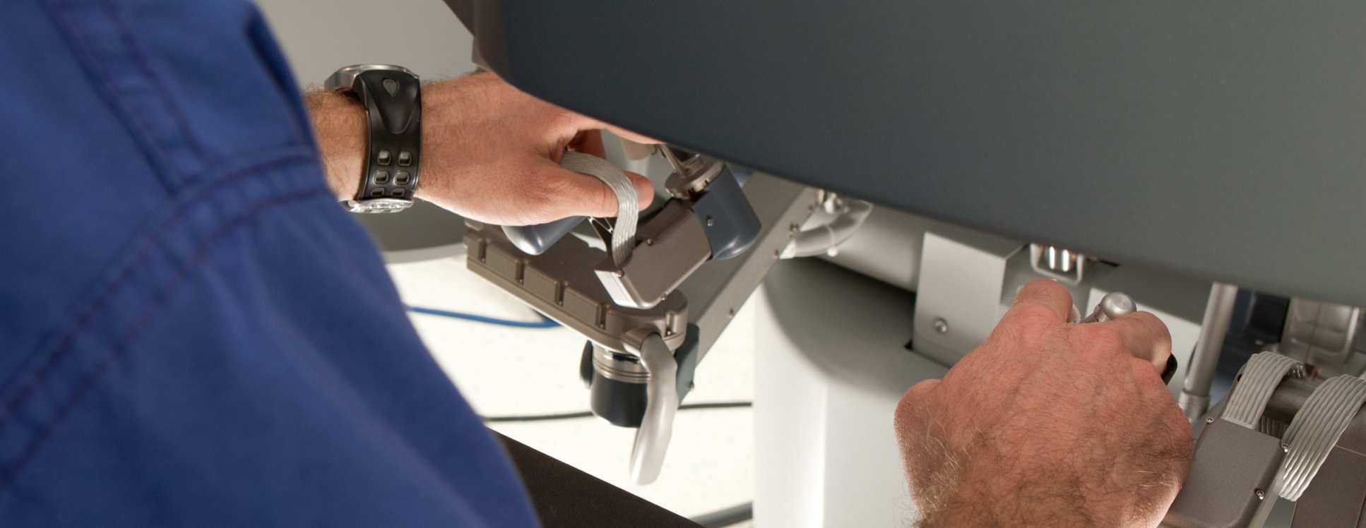
Imperial was the site for the installation of the world’s first robotic endovascular catheter for use in the vascular tree. Our group has published first in-vitro evidence of the advantages of robotic catheter systems for endovascular treatments in terms of operator performance, safety, overcoming difficult anatomy and learning of skills. The world’s first robotic endovascular aortic aneurysm repair was performed at the St Mary’s site receiving National and International press attention and providing proof of the concept of robotic endovascular therapy. The development of a specific robotic vascular catheter has been guided by the research from our site and input from our group, acknowledged by our industry associates. The world’s first vascular specific robotic catheter system was installed for clinical use in 2012 at Imperial. This system has now been used in 150 cases and the safety and efficacy of the system addressed. The robotic system has now been installed in over 15 sites. Further studies are ongoing to assess the advantages of this robotic system in different procedures.
Researchers
Helix is a joint collaboration between Imperial College London and the Royal College of Art. Our multi-disciplinary team is uniquely placed on the frontline of healthcare, to address real healthcare problems by translating research into evidence-based solutions. We use various design methods to rapidly dissect problems, envisage opportunities and prototype solutions.
Related centres
Researchers
HARMS Lab: Human-centred Automation, Robotics and Monitoring for Surgery
The HARMS lab employs patient and surgeon-specific approaches, perceptually-enabled methodologies and frugal innovation through human-centred design to deliver enabling and disruptive solutions in the areas of monitoring, automation and robotics for surgery. Our human-centred paradigm starts with the people we are designing for and ends with new solutions that are tailored to suit their needs.
Find out more
Researchers
The image-enhanced operating environment defines a new paradigm for information-rich surgical workflow, and brings the broadest range of modalities to bear on key surgical procedures. Novel intraoperative surgical imaging techniques play a central role, enabling the identification of targets at a disease-specific level. The long-term strategy addresses important research questions to ensure that the right information is delivered at the right time, through interfaces most appropriate to the intervention.
In the largest study of its kind, our platform has demonstrated a clinical subjective efficacy, and objective non-inferiority, over the status quo for robot-assisted partial nephrectomy. The following projects are also ongoing:
1. Integration of Image Guidance in Soft Tissue Minimal Access Surgery
This project seeks to understand how image guidance has been adopted in clinical practice, and in what ways it has been integrated within current surgical workflows.
2. Image Guidance for Predicting the Functional Outcome for ‘Salvage’ Resection in Pelvic Cancer Patients
This study focuses on the use of multimodal image analysis to assess the effect of radiotherapy on the urethral sphincter complex. Using intraoperative imaging guidance platforms, this study aims to facilitate precise resection with concomitant preservation of function, reducing the need for further treatment of incontinence.
3. Improved Visualisation for Fistula Repair Surgery
This trial seeks to apply the existing image guidance platform to certain perianal interventions. Specifically, in anal fistula surgery, improved visualisation of the anal sphincter complex and fistula tract will allow complete excision of fistula tissue whilst minimising damage to the sphincter complex. Several display platforms suitable for open surgery are under evaluation.
4. Autonomous Ultrasound-Guided Tissue Dissection
Using robotic partial nephrectomy as an index procedure, and employing PVA cryogel tissue phantoms in a reduced dimensionality setting, this study sets out to achieve autonomous tissue dissection with a high-velocity water jet under ultrasound guidance. The open-source da Vinci Research Kit (DVRK) provides the foundation for a novel multimodal visual serving approach, based on the simultaneous processing and analysis of endoscopic and ultrasound images.
Our researchers
- Mr Erik Mayer
- Dr Philip Pratt
- Mr Justin Vale
- Mr Neil Tolley
- Professor Zoltan Takats
- Mr James Kinross
- Mr Alex Von Roon
- Mr George Mylonas
- Mr James Dilley
- Mafalda Camara
- Ismail Omar
- Philip Pucher
Intelligent surgical devices combine surgical dissection techniques with mass spectrometric analysis of the by-products (smoke, liquefied tissue) of the dissection for enhancing real-time tissue identification for surgical decision making. It has been applied for multiple cancer types and a prospective study demonstrated 97.75% correct classification. Work is performed with support from Waters and ERC.
The performance testing of iKnife for margin control is in progress. The technology provides data on tumour phenotype including molecular features for stratifying oncological therapies (e.g. ER or KRAS status). We will deploy this technology in prospective trials to assess its ability to improve reoperation rates. We are also combining the technology with computer assisted surgery and developing minimally invasive applications for screening, diagnostics and precision polyp phenotyping through our endoscopy program.
Chemical endoscopy: We are developing REIMS based technologies for the real-time assessment of large polyps and other lesions of the GI tract. The technology allows for not only the in situ real-time identification of tissues, but also the characterisation of the host-associated microbiome, which has significant relevance with regard to the early-stage detection of potentially malignant gastrointestinal diseases. We are planning to launch multicentre randomised prospective trials in the next 24 months.
Chemical / spectroscopic real-time augmented histology: The in situ mass spectrometric imaging (MSI) was developed at Imperial using Desorption Electrospray Ionization (DESI). This technique permits a new layer of molecular information to be overlaid onto histopathological images and opens up exciting new avenues in molecular pathology. Briefly, most phenotypic information necessary for proper patient stratification can be obtained in a single step of untargeted MSI analysis, potentially replacing a multi-step histological workflow. We have developed bioinformatic processes for studying spatially resolved chemical perturbations in cancer. The method also provides a novel approach for pharmacometabonomic analyses in cancer tissues. We will trial the technique in breast, ovarian, colorectal and brain cancers to further assess its diagnostic accuracy and clinical potential. Furthermore, we are combining MSI and iKnife approaches in order to fully utilise the capabilities of MS-based tissue analysis technologies.
Find out more about the iKnife and REIMS technology.
Collaborators
Researchers
This work takes place collaboratively with the neuroergonomics and perception laboratory and Robot Brain Interaction Group, Hamlyn Centre at Imperial College London.
Related centres
Our research is based around the development and application of photonics technology with endoscopy for surgical imaging applications, including multispectral imaging, polarization-resolved imaging, fluorescence imaging, and the use of fluorescently labelled gold nanorods for theranostics. Further projects include work on the development of illumination and vision systems for endoscopy combining miniature light sources such as LEDs and laser diodes with computer vision techniques for structured lighting and tissue surface reconstruction as well as the use of robotic guidance of optical probes. These devices are finding application in minimally invasive surgery and in the development of new flexible robotic-assisted surgery systems.
Prospective feasibility study of LED light and fluorescence cystoscopy
A new endoscopic light source has been developed by the research team proposing this trial that is based on LED technology. The aim of this study is to elucidate the feasibility of using a multicoloured LED (light emitting diode) light source in endoscopy (LED Light) as an alternative to the standard Xenon white light found in STORZ system, which is known as 'Blue Light'. The LED Light is a safe and reliable light source that has a number of advantages over the xenon lamp, including the control of the white light colour balance, as well as usability and cost improvements.
Real-time tissue validation using NIR fluorescence imaging
The purpose of this study is to investigate whether a novel affordable and compact fluorescence sensitive imaging system (FSI) can be used to visualise subtle differences between healthy and cancerous tissue. This technique can also be used in breast reconstructive surgery to monitor the vasculature system and tissue oxygenation. It is hoped that the results of this study can be used in the future to help clinicians detect and evaluate the cancerous tumour and its boundaries, hence preventing unnecessary tissue resection. Furthermore, the outcome of this research may help increase the chance of successful reconstructive surgeries and therefore avoiding additional operations.
Researchers
Our approach is to develop surgical robots that expand the previous generation of laparoscopic devices and to improve intrasurgical monitoring technologies and methods to characterise the impact of the intervention. The Hamlyn Centre was established for developing safe, effective and accessible technologies that can reshape the future of healthcare for both developing and developed countries. The Hamlyn Robotics Centre focuses on technological innovation with a strong emphasis on clinical translation, resulting in direct patient benefits with global impacts.
This research includes the design and evaluation of laparoscopic instrumentation and imaging systems and the use of Human Reliability Assessment (HRA) techniques in the clinical environment to identify committed errors and study performance shaping factors.
Related centres
Our researchers
Key researchers in surgical technology
Professor Paul Abel
/prod01/channel_3/media/migration/faculty-of-medicine/Abel,-Paul--tojpeg_1487169719978_x4.jpg)
Professor Paul Abel
Professor of Urology
Mr Colin Bicknell
/prod01/channel_3/media/migration/faculty-of-medicine/Bicknell,-Colin--tojpeg_1487169434784_x4.jpg)
Mr Colin Bicknell
Clinical Senior Lecturer
Professor Ara Darzi
/prod01/channel_3/media/migration/faculty-of-medicine/Darzi--tojpeg_1487688347952_x4.jpg)
Professor Ara Darzi
Professor of Surgery
Professor Dan Elson
/prod01/channel_3/media/migration/faculty-of-medicine/Elson,-Dan--tojpeg_1487170365258_x4.jpg)
Professor Dan Elson
Professor of Surgical Imaging
Dr Dominic King
/prod01/channel_3/media/migration/faculty-of-medicine/King,-Dominic--tojpeg_1487171304745_x4.jpg)
Dr Dominic King
Honorary Clinical Lecturer
Mr James Kinross
/prod01/channel_3/media/migration/faculty-of-medicine/Kinross,-James--tojpeg_1487171386744_x4.jpg)
Mr James Kinross
Clinical Senior Lecturer in Colorectal Surgery
Mr Daniel Leff
/prod01/channel_3/media/migration/faculty-of-medicine/Leff,-Richard--tojpeg_1487171759314_x4.jpg)
Mr Daniel Leff
Clinical Senior Lecturer in Breast Surgery
/prod01/channel_3/media/migration/faculty-of-medicine/Lo,-Benny--tojpeg_1487171819675_x4.jpg)
Mr Erik Mayer
/prod01/channel_3/media/migration/faculty-of-medicine/Mayer,-Erik--tojpeg_1487171868573_x4.jpg)
Mr Erik Mayer
Clinical Senior Lecturer
Dr George Mylonas
/prod01/channel_3/media/migration/faculty-of-medicine/Mylones,-George--tojpeg_1487172467772_x4.jpg)
Dr George Mylonas
Lecturer in Robotics and Technology in Cancer
Dr Philip Pratt
/prod01/channel_3/media/migration/faculty-of-medicine/Praqtt--tojpeg_1493723904325_x4.jpg)
Dr Philip Pratt
Research Fellow
Professor Zoltan Takats
/prod01/channel_3/media/migration/faculty-of-medicine/Takats,-Zoltan--tojpeg_1487172950779_x4.jpg)
Professor Zoltan Takats
Professor of Analytical Chemistry