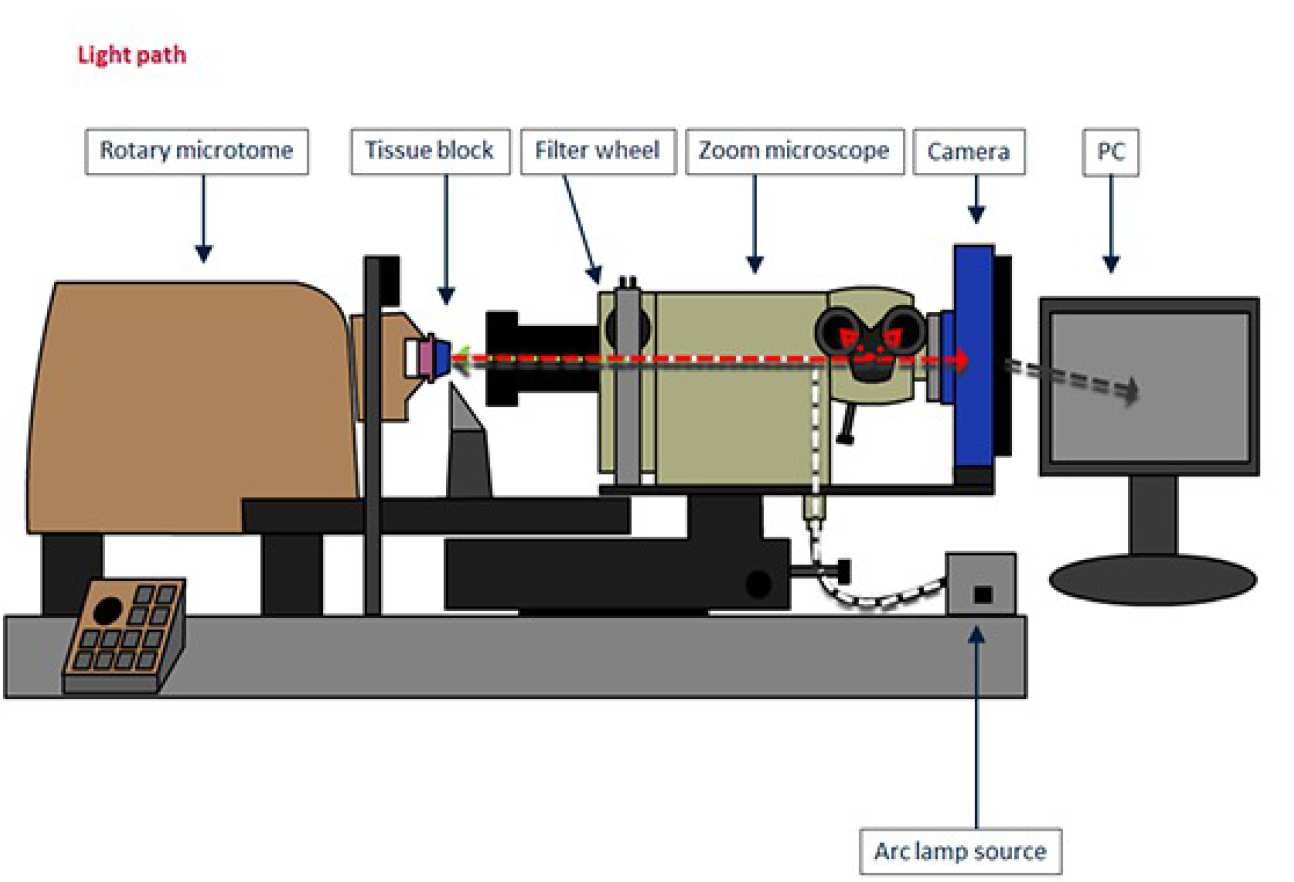
The diagram below shows the path of light through the Histocutter, with each of the main parts labelled:
How does it work?
This Histocutter is a robotic 'cut and view' 3D imaging system. Samples are infiltrated with opaque dyes (to block the out-of-focus light), and embedded in wax or hard plastic, before being mounted in the chuck of a rotary microtome.
When a cutting session is initiated, the microtome cuts a predefined slice from the block, and then the cut block face is imaged using a fluorescent microscope and high resolution camera. Up to four images can be taken for each slice, each with a different fluorescent channel, by means of the automated filter wheel attached to the micoroscope.
The images are then automatically sent to a computer, where they are aligned and stored (in duplicate). The process is repeated until the desired depth of block is cut. The entire process is automated, using a bespoke control software program.
All the cutting and imaging parameters are stored in a text file with the folder containing the images. The system outputs a high resolution aligned multi-channel stack of images, which can be loaded straight into a 3D visualisation package, such as
- imageJ (https://imagej.nih.gov/ij/)
- and ITKsnap (http://www.itksnap.org/pmwiki/pmwiki.php)
- Amira
- Volocity 3D
for reconstruction and segmentation of data obtained with the histo-cutter.
Samples types
Organs, scaffolds, biopsies, cell pellets, and other histologic or material based samples.
The criteria is that they must be smaller than 6 x 6 mm (if you want to see the entire sample) and able to be cut on a typical histology wax knife or using a tungsten carbide knife. So bone, hydroxyapatite, soft tissue etc is fine, but no glass or metal samples and we cannot do cryo.
Staining
Samples may be stained ‘en bloc’ or perfused with dyes, injected with growth markers or peptides, or simply imaged using their native autofluorescence. In addition, reporter genes such as GFP or RFP can be imaged.
Embedding
Samples may be embedded in MMA (methyl methacrylate), Histology wax, low temperature polyester wax, or Epon/araldite resin. The choice depends on which type of tissue or material one is trying to visualise, and the type of labels the tissue contains.
Sample preparation
Staining
- Native tissue (autoflorescence)
- En-bloc staining with dyes
- Perfusion with dyes
- Reporter genes (e.g. GFP)
- Injected growth markers
- Injected ECM peptides
Embedding media
- NMA (methyl methacrylate)
- Histology wax
- Epon/araldite?
- No cryo in this system
Opaque dyes used to infiltrate sample and embedding media, in order to block out-of-focus fluorescence.