


JEOL JEM-2100F TEM
The TEM is routinely used for characterising the microstructure at interfaces in ceramics, Metals and Biological samples. Morphology and distribution of nano-size particles, lattice imaging and chemical analysis can be studied.
The TEM is computer-controlled allowing users to record operation conditions, recall stored sample grid positions and perform low-dose imaging. This is useful for examining biological samples that are sensitive to the high-energy electron beam. To record high-resolution images using less energetic beams, the instrument is fitted with a Gatan Orius SC 1000 camera (2×4k) GIF Ultras-can camera (2k×2k), high-angle annular dark field detector, a Gatan annular dark field detector/bright field detector, as well as a Gatan Quantum image filter (GIF) system.
Further chemical analysis can be carried out on the Oxford Instruments INCA/Aztech EDS 80 mm X-Max detector system, which is capable of light-element (Z>5) and can be combined with STEM for nanometre spatial resolution.
Au nanoparticles and Convergent beam diffraction pattern.
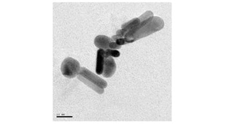
Au nanoparticles and Convergent beam diffraction pattern 1
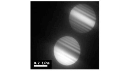
Au nanoparticles and Convergent beam diffraction pattern 2
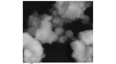
STEM Image
Images JEOL JEM 2100F - bottom
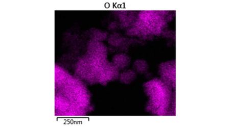
Oxygen EDX Map
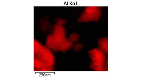
Aluminium EDX Map

Silicon EDX Map
JEOL JEM-2100F TEM help and support
-
Dr Mahmoud Ardakani
/prod01/channel_2/media/migration/faculty-of-engineering/Mahmoud-Ardakani--(Nov-2013)--tojpeg_1499781242792_x4-6.jpg)
Personal details
Dr Mahmoud Ardakani Research Officer, Harvey Flowers Electron Microscopy SuiteSend email+44 (0)20 7594 6739
Location
Department of Materials
Royal School of Mines
Lower Ground Floor, LG05