Our brain is the most complex system known to us. Road traffic incidents, sporting activities, falls, disease or surgical interventions can subject our brain to large mechanical loads, which can damage our brain.
At HEAD lab, we focus on understanding the effects of mechanical loading on the brain and using this fundamental understanding to develop applied solutions to predict and prevent brain injuries.
Our Projects
We develop high fidelity computational models of brain biomechanics and use them to predict the effects of mechanical loading on brain tissue.
We have developed the Imperial College Finite Element Model of Brain Injury Biomechanics, which incorporates fine details of brain anatomy, such as sulci.
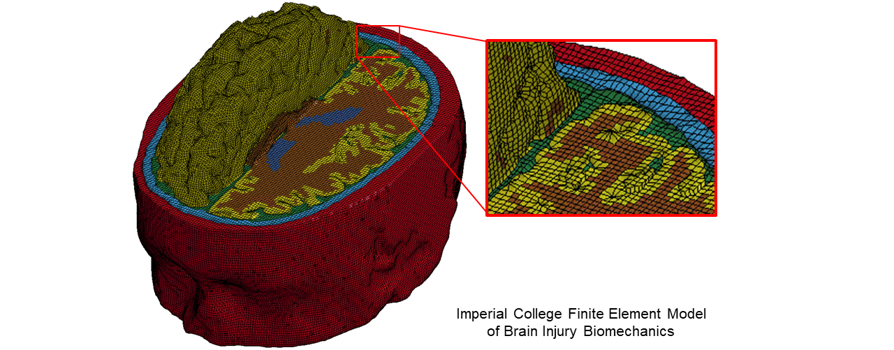
This model has allowed us to predict the location of the pathology in the dementia disease, chronic traumatic encephalopathy (CTE). Press release: https://www.imperial.ac.uk/news/179073/3d-model-american-football-players-brain/
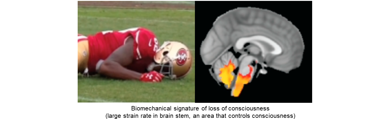
In addition, this model has also allowed us to reveal the biomechanical signature of loss of consciousness. Press release: https://www.imperial.ac.uk/news/243996/sports-related-brain-injuries-reveal-mechanics/
Biomechanics of Axonal Injury
Exposure of the white matter to loading can damage its key structural elements, the axons. Axons are the long projections of nerve cells. Axonal damage can determine long-term outcomes after traumatic brain injuries. Our aim is to better understand how deformation of the white matter in trauma and disease can damage axons. We use this understanding to improve our prediction tools and design prevention strategies.
This work is funded by a Dyson School of Design Engineering PhD Scholarship. We collaborate with neuroscientists at Imperial College Brain Sciences Department to access human data and understand injury patterns.
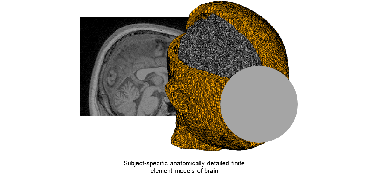
Individualised Brain Models
Biomechanical brain models are built based on a geometrical description of the brain anatomy, obtained from brain images such as MRI. We have developed a semi-automatic pipeline that allows us to build a brain model from an MRI scan, incorporating fine details of the brain anatomy. Our aim is to use this approach to improve our understanding of the effects of anatomy on the mechanical response of the brain in trauma and disease. We will incorporate these findings in brain surveillance systems.
This work is part of the Sports and Wellbeing/Cellbond Impact Solutions/Royal Academy of Engineering Senior Research Fellowship.
From Biomechanics to Pathology
This work is another example of the strong and unique collaboration between Design Engineering and Brain Sciences at Imperial College. We used high fidelity biomechanics modelling of traumatic brain injury in rats, empirical modelling of controlled cortical impacts in rats, high intensity MRI, and quantitative histopathology to understand the relationship between white matter deformation and damage. We showed that large brain deformations produced at the time of injury can predict structural changes in white matter two weeks post-injury. This understanding confirmed the validity of using brain tissue deformation to predict short and long-term effects of mechanical loading on brain tissue. Press release: https://www.imperial.ac.uk/news/212873/precise-mapping-shows-brain-injuries-inflict/
This work was funded by Wellcome Trust Networks of Excellence and Centre for Blast Injury Studies.
Biomechanics of Vascular Injury
Cerebral vasculature injury is a form of traumatic brain injury (TBI), where the blood vessels in the brain are damaged, causing intracranial bleeding. We have led studies to understand the relationship between mechanical loading of vessels and their damage in humans and rats.
We have incorporated details of the venous system within our brain model. With this model, we studied the patterns of large strains in small vessels during head loading and the location of microbleeds after head loading. We found an overlap between large stretches of vessels and microbleeds in a rugby impact. Our aim is to extend this study to a range of cases and establish a threshold for vascular damage.
An overview of our paper on prediction of blood brain barrier damage by biomechanical finite element modelling of vascular injury

We have also incorporated a map of cerebral vasculature, with a 5 microns resolution obtained from synchrotron imaging, in our finite element model of rat brain biomechanics. Using this model and quantitative histopathology of rat brain tissue subjected to impacts, we found that there is an interaction between the anatomy of capillaries and loading distribution in producing blood-brain barrier damage. Press release: https://www.imperial.ac.uk/news/226433/brain-injury-computer-models-brain-blood
An overview of our paper on prediction of blood brain barrier damage by biomechanical finite element modelling of vascular injury

This work was funded by Wellcome Trust Seed Awards in Science and EPSRC DTP studentship.
We develop technologies to estimate mechanical forces applied to the head and brain in sporting and road traffic incidents, providing objective information for a range of applications that require brain exposure monitoring.
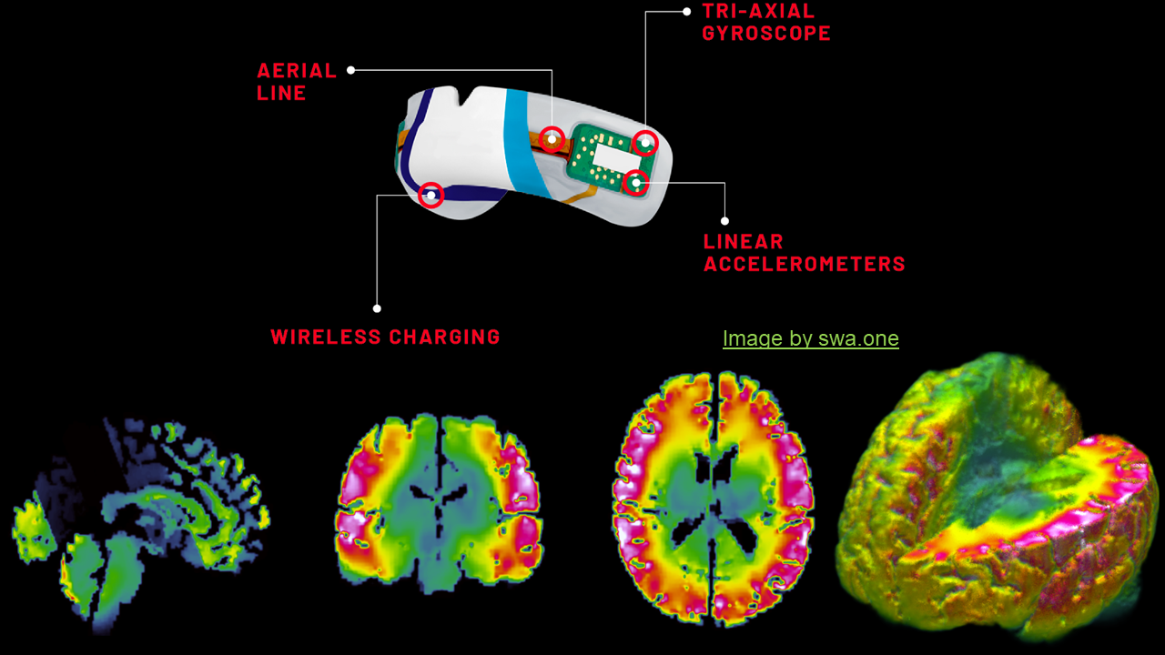
Monitoring brain biomechanics in sports
One key application is in sporting, where these technologies can be used to measure biomechanical forces that a player has experienced in a match, season or their career. This information can benefit medics, coaches, parents and guardians, and sport governing bodies.
Instrumented mouthguards are mouthguards that are equipped with miniature sensors that measure translational and rotational motion. They can measure translational and rotational motion of the head during head acceleration events. We collaborate with sport governing bodies and mouthguard companies to access thousands of such data to enable our research.
We develop fast running surrogate models for our detailed brain models to predict brain loading in a fraction of a second. Machine learning has been used to build such models. Our aim is to develop novel surrogate models that can address practical problems and work towards their implementation and application in brain health surveillance systems.
This work has been funded by Sports and Wellbeing Analytics/Cellbond Impact Solutions/Royal Academy of Engineering Senior Research Fellowship and MRC TBI-REPORTER.
AutoTriage: brain injuries in road traffic incidents
Traumatic brain injury is one the most time-sensitive injuries that road traffic casualties sustain. Rapid and tailored post-crash response can considerably reduce risk of death and long-term consequences.
We have shown that on-board vehicle sensors can be used to predict the severity of traumatic brain injuries in road traffic incidents. Large-scale data-driven analysis of the UK's Road Accident In-Depth Study (RAIDS) database has enabled us to determine this relationship. We are now extending this work to improve the prediction algorithms and to implement such system in medical emergency responses.
The combined expertise between the Transport Research Laboratory's automotive Engineering, Safety & Technology sector and Imperial College London's Medical & Dyson Design Engineering Schools allowed for a unique approach to improving the clinical outcome for road users involved in Road Traffic Collisions. Press release: https://www.imperial.ac.uk/news/233900/road-accident-data-could-help-predict/
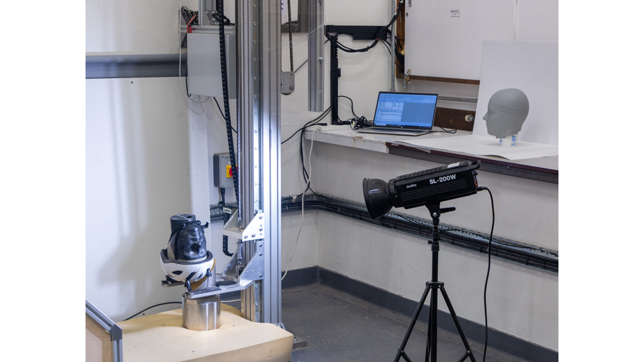
Introducing HEAD Lab's Helmet Test Facilities
This work has been led by Dr Claire Baker with funding from Transport Research Laboratory, EPSRC Centre for Doctoral Training in Neurotechnology, EPSRC Doctoral Prize Fellowship and Road Safety Trust.
Helmets are designed to prevent head and brain injuries. Their protection is assessed according to standard test methods, but helmet standards have not been updated for decades. Hence, they do not reflect our improved understanding of brain injury biomechanics and injury causation.
Our research on helmets focuses on developing helmet test methods that accurately represent real-world scenarios and predict risk of different types of brain injuries. We actively pursue approaches to disseminate the findings of our research among a range of stakeholders, including consumers, manufacturers, standard committees and authorities.
Hiper: cycle helmet rating
Cyclists who choose to wear a helmet do so to protect their head if they are involved in an incident, such as falls or collisions. In the absence of objective information to help them choose a safer helmet, they may rely on claims made by manufacturers or the helmet price.
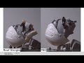
To address this challenge, we have developed Hiper, a cycle helmet rating system, based on advanced brain injury biomechanics research and extensive evidence from real-world incidents. We shortlisted 30 most popular helmets used by the UK cyclists and evaluated their performance using HEAD lab’s helmet test rig. We have published the results under the Hiper brand and through a dedicated website, developed for consumers.
Our aim is to continue the rating and extend it to other types of helmets. This can be achieved through an independent organisation that manages the testing, dissemination and research in collaboration with other stakeholders.
This work was funded by the Road Safety Trust and Innovate UK. The Road Safety Trust has provided further funding under their large grants scheme to continue the rating and extend it to Children’s helmets.
Towards rating industrial helmets
Industrial helmets, or hardhats, are widely used in industrial settings to prevent head and brain injuries. Current standards are designed to test protection against falling objects but falls and trips are the dominant cause of traumatic brain injuries in industrial places.
We have determined head impacts conditions in falls and trips using computational modelling of over 1,600 falls and trips. We resorted to modelling due to the unavailability of real-world data in this field. The simulation results have informed a new test protocol for assessing hardhats. We have now tested a range of industrial helmets under these conditions, showing that the tests can distinguish between these helmets. Our aim is to extend this work by developing a rating system for industrial helmets and disseminate the results under the Hiper brand.
Introducing HEAD Lab's Helmet Test Facilities
This work has been funded by the Innovate UK and MIPS. We are seeking further funding to continue this work for the benefit of workers exposed to workplace hazards.
Assessing facial impacts in motorcycle incidents
Impacts to the face in cycle, motorcycle and e-scooter incidents can cause brain injuries and complex fractures, such as facial and basilar skull fractures, with life-changing and fatal consequences. However, apart from full-face helmets, there is no effective solution for protection against facial impacts. In addition, the existing standard methods for testing facial impacts use linear acceleration of the head or deflection of the helmet chin-bar to assess the chin-bar of full-face helmets. This approach does not address protection against facial injury and particularly basilar skull fracture which is caused by the neck forces.
Our aim is to bridge this important gap by designing new methods for testing face protection solutions. We are using in-depth road traffic collision databases to better understand the consequences of facial impacts and determine representative conditions for developing new test methods.
This work is funded by Autoliv Research and involves collaboration with trauma experts at Imperial College St Mary’s major trauma centre.
Other projects on head protection
HEAD lab has led several other projects on head protection. More information can be found in the news releases below.
E-scooter falls: https://www.imperial.ac.uk/news/233378/e-scooter-simulations-highlight-head-injury-risk/
This work was cited by PACTS: https://www.pacts.org.uk/wp-content/uploads/PACTS-The-safety-of-private-e-scooters-in-the-UK-Final-Report.pdf
Sikh Turbans: https://www.imperial.ac.uk/news/251115/turban-style-thickness-affects-head-injury/
Cycle helmets: https://www.imperial.ac.uk/news/221193/new-testing-bike-helmets-accounts-head/
Looking for a collaboration?
We have been collaborating with several industries across the world in various capacities; for example consultancies, Innovate UK projects, and direct funding. Please reach out via the email on the left side of this page to discuss.
We are always looking for students with excellent academic profiles, who are passionate about translational biomechanics research. Please reach out to discuss. In your email, it is important to explain why you want to study at the HEAD lab.

Contact us
For queries, please contact Mazdak Ghajari:
- Email: m.ghajari@imperial.ac.uk
- Phone: +44 (0)20 7594 9236
Opportunities
If you are interested in the work we do and are interested in postgraduate (PhD) or postdoctoral research, you can find more information about scholarships and fellowships below:
- General Scholarship Information
- China Scholarship Council
- President's PhD Scholarships
- Imperial College Research Fellowships
We are also interested in Undergraduate Research Opportunities Programme applicants.
Please get in touch with Mazdak Ghajari in the first instance if interested in applying.