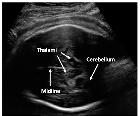
 Intrapartum ultrasound in second stage of labour: determining prognostic factors for type of birth. We invite mothers to take part in our study that are having their first baby at term (37-42 weeks) and wish to have a vaginal (normal) delivery. We use ultrasound to assess which way the baby is positioned and also how low the baby’s head is in the birth canal. When the healthcare practitioner confirms the cervix (neck of the womb) is fully dilated (10 cm), we perform the ultrasound scan at this time. There are two ultrasound methods: transabdominal and transperineal. Transabdominal ultrasound scan is similar to scans that are done routinely in pregnancy, assessing the baby's position and function of the placenta.
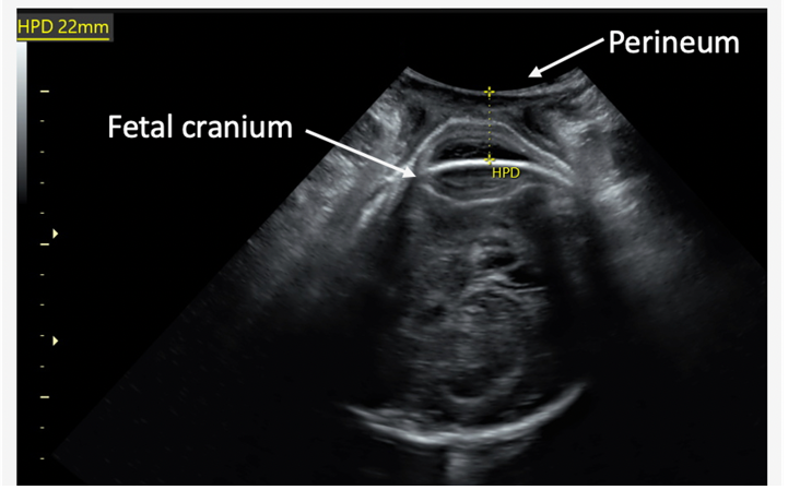 Transperineal scanning involves placing a covered and clean probe between the labia (outside the vagina) for 1-2 minutes without undue pressure, assessing the baby's descent in the birth canal. Both the transabdominal and transperineal scans are performed as part of this study. The total scan time is 5 minutes and routine care in labour is not affected. Information from the ultrasound scans may help us to predict whether a caesarean section or assisted vaginal birth will be needed and thus may help guide women and birth attendants in the future.
Researchers
Dr Mariya Kovalenko
/prod01/channel_3/media/images/people-list/Dr-Mariya-Kovalenko.png)
Dr Mariya Kovalenko
Clinical Research Fellow