Researchers discover how viruses such as HIV unravel human DNA

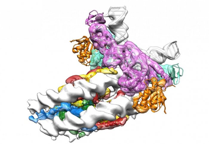
Cryo-electron microscopy structure of a retroviral integrase attacking a human nucleosome.
An international team of researchers has discovered important new details of how retroviruses such as HIV take over healthy cells.
The research, led by scientists from the Francis Crick Institute and Imperial College London and published today in Nature, may eventually lead to more effective treatments to suppress viruses in people who have been infected.
Retroviruses insert their genomes into the chromosomes of host cells, an irreversible process which makes them particularly difficult to eradicate. The viral enzyme responsible for this process is called integrase and it is carried by every retrovirus. Using a very powerful electron microscope, the research team discovered how integrase captures tightly-wound packages of human DNA called nucleosomes, in order to insert the viral DNA.
Professor Peter Cherepanov, from the Francis Crick Institute and Imperial College London, said: “Cellular DNA is tightly packaged in the form of nucleosomal arrays, with each nucleosome acting as a tiny reel. This compaction could be expected to present an obstacle for viral integration. In the course of this study, we were able to explain how the virus captures and opens up human nucleosomes to insert its DNA into a human chromosome.
“The more we learn about the rules of engagement between the viral integration machinery and host cells, the easier it will become for us to break the chain of events that lead to persistent infection. This can lead to more effective antiretroviral drugs to treat HIV infection.
“There is also a potential benefit for gene therapy. The unique ability of retroviruses to efficiently integrate their genetic material into host cell chromosomal DNA means they could be used to deliver highly targeted drugs as part of gene therapy. However, uncontrolled integration by retroviruses carries the risk of serious side effects. Understanding how to selectively direct integration will potentially aid the development of safer gene therapy.”
Another Francis Crick Institute researcher on the study, Alessandro Costa, explained how the team visualised the interaction between integrase and a nucleosome. “Starting from 2D images of individual molecular assemblies, we have reconstructed a high-resolution 3D view of integrase attacking a tiny fragment of a human chromosome, 10 billionths of a metre in diameter. This achievement, unconceivable only four years ago, was made possible by using a very sophisticated electron microscope, new generation cameras and innovative software for data analysis,” he said.
“Biological nanomachines such as integrase can now be imaged in unprecedented detail as they perform their work. Thanks to these new tools we can rethink the way we ask scientific questions”.
Sir Paul Nurse, director of the Francis Crick Institute, said: “This is a fascinating and important discovery which increases our understanding of the way retroviruses work. It has important long-term implications, not only for leading to improvements in HIV treatments but also for treating a range of other diseases which may benefit from gene therapy.”
The research team included scientists from Imperial College London, the Dana-Farber Cancer Institute in Boston, the Gorlaeus Laboratory in Leiden, and TU Dresden.
The work was supported by funding from the European Commission and National Institutes of Health.
Reference: D.P. Maskell et al. 'Structural basis for retroviral integration into nucleosomes.' Nature 2015. doi: 10.1038/nature14495
Article text (excluding photos or graphics) © Imperial College London.
Photos and graphics subject to third party copyright used with permission or © Imperial College London.
Reporter
Peter Zarko-Flynn
Communications and Public Affairs