The brains of alcohol dependents and binge drinkers may recover differently

Cells that clear damage in the brain are less active in alcohol-dependent patients after withdrawal than in models of adolescent binge-drinking.
People who have become alcohol-dependent suffer symptoms of withdrawal while abstaining from drink. These include cognitive impairment, such as memory problems, making plans and being able to act flexibly.
Previous evidence from research in mice has suggested that these impairments might be caused by the over-activity of cells called microglia. Microglia maintain the brain by removing damaged neurons and synapses and fighting infections.
However, excess microglia activity can be harmful, producing chemicals that cause collateral damage to healthy nerve cells. This process is implicated in brain disorders such as Alzheimer's and Parkinson’s diseases.
Microglia activity has previously been found in experiments to increase after mice are given high doses of alcohol which make them suffer from withdrawal symptoms. This led researchers to ask whether the same damaging process was occurring in middle-aged alcohol dependents suffering withdrawal.
Finding the opposite of what was expected
To investigate, a team led by Imperial College London and Kings College London scanned the brains of alcohol-dependent patients and healthy volunteers looking for microglial activity.
Previous knowledge suggested that we would find greater microglial activity in alcohol-dependent patients’ brains, but in fact we found the opposite.
– Dr Nicola Kalk
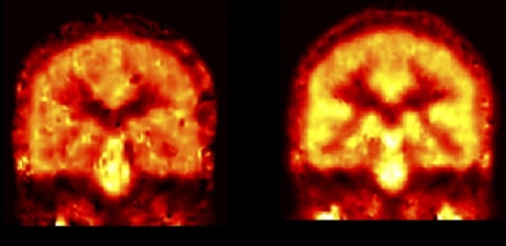
What they found was the opposite of what they expected. The experiment revealed that alcohol-dependent patients abstaining from alcohol actually show less microglial activity than healthy volunteers.
They also found that in healthy people, the greater the levels of microglial activity in the hippocampus, the better their memories were.
The team believe the discrepancy between the mouse models and the human subjects might be because mouse models of alcohol abstinence better represent adolescent binge-drinkers, rather than middle-aged alcohol dependents.
The results point to a different role for microglial activity. The team suggest that in adolescent binge-drinking mice, the surge of microglial activity may help to restore the hippocampus, the area of the brain involved in memory.
In the brains of human alcohol-dependent patients, this activity is dampened, either because the microglia are not functioning properly or because they have been lost.

Image caption: Microglial activity in alcohol-dependent brain (L) and control brain (R)
The team’s result supports other research that shows that microglial over-activity might not always be harmful, and that there might be two types of activity that either cause damage or cause repair.
Study author Dr Nicola Kalk, from the National Addictions Centre at King’s College London and the Division of Brain Sciences at Imperial, said: “Previous knowledge suggested that we would find greater microglial activity in alcohol-dependent patients’ brains, but in fact we found the opposite.
"Combined with our other finding – that microglial activity was associated with better memory in healthy volunteers – this led us to consider the idea that microglial activity can sometimes have a positive effect on cognitive function that is lost in alcohol-dependent patients.”
This is an important study to help us understand what processes are involved in the damaging effects of alcohol on the brain.
– Professor Anne Lingford-Hughes
The researchers want to explore possible interventions to boost microglia activity in alcohol-dependent patients, and also the mechanism that seems to confer an advantage to young binge-drinking brains, allowing them to avoid the cognitive decline seen post-drink in dependent brains.
To determine microglial activity, the team compared the brain scans of healthy volunteers and patients with alcohol dependence. They used a technique called positron emission tomography that allowed them to quantify the microglial activity in each volunteer’s hippocampus. The results of the study were published this week in the journal Translational Psychiatry.
Professor Anne Lingford-Hughes, from the Centre for Psychiatry at Imperial, said: “This is an important study to help us understand what processes are involved in the damaging effects of alcohol on the brain. Without this knowledge we will not be able to develop treatment strategies to improve brain recovery to optimise cognitive function.”
-
‘Decreased hippocampal translocator protein (18 kDa) expression in alcohol dependence: a [11C]PBR28 PET study’ by N J Kalk, Q Guo, D Owen, R Cherian, D Erritzoe, A Gilmour, A S Ribeiro, J McGonigle, A Waldman, P Matthews, J Cavanagh, I McInnes, K Dar, R Gunn, E A Rabiner and A R Lingford-Hughes is published in Translational Psychiatry.
Article text (excluding photos or graphics) © Imperial College London.
Photos and graphics subject to third party copyright used with permission or © Imperial College London.
Reporter
Hayley Dunning
Communications Division