Imperial and Leiden University collaborate on world-leading AR assessment

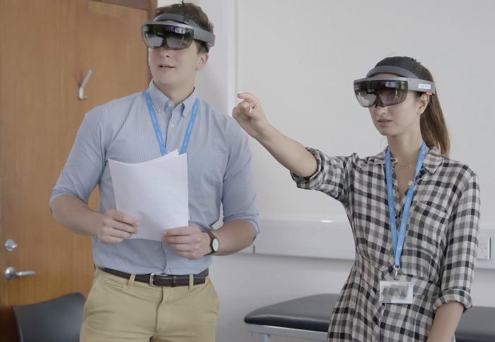
Postgraduates testing Microsoft HoloLens AR headsets
School of Medicine students are trialling Augmented Reality (AR) headsets, bringing anatomy education out of textbooks & closer to real-world practice
The pioneering trial involving MBBS students, the College's Digital Learning Hub, and Leiden University Medical Center, is exploring whether AR technology could be introduced into medical student examinations.
AR headsets are an increasingly common feature of training courses for graduate medics, and are beginning to appear more frequently within the curricula of undergraduate programmes. However, no higher education institutions are yet using this technology for student assessment. This means that although students are impressed by the potential of AR during their time as undergraduates, they are still faced with a traditional exam paper when they reach the end of their studies.
 For Dr Amir Sam (left), of Imperial's Department of Medicine, this is a needlessly disjointed way of training the medics of the future. Dr Sam said: "Having reviewed the assessment frameworks of a number of other universities and professional bodies, I've seen no one else try to do what we are doing. It really does seem to be a first.
"Introducing this technology means we can test students' ability to spatially orientate themselves when interacting with a digital version of the human anatomy. It really elevates the quality of our assessment and encourages far deeper understanding than simply looking at anatomy on paper."
"AR has the potential to replicate...scarce resources in a much more sustainable way. With this technology, anatomists will reduce the need to use cadavers and centuries-old dissections in the teaching and assessment of human anatomy." Dr Amir Sam
In his role as Head of Curriculum & Assessment Development for Imperial's School of Medicine, alongside assessment roles with a number of UK and European medical schools, Dr Sam has a bird's-eye-view of the profession. He said: "We are all acutely aware of resource limitations within healthcare. This, coupled with an increasing number of medical students makes conducting valid and reliable assessments particularly difficult, especially when assessing resource-intensive disciplines, such as anatomy.
"AR has the potential to replicate some of these scarce resources in a much more sustainable way. With this technology, anatomists will reduce the need to use cadavers and centuries-old dissections in the teaching and assessment of human anatomy."
Visualising medicine
Student Fellow, Adam Misky, was involved in a number of stages of the trial. He participated in the initial design phase and served as an assessor both when trialling the assessment on a panel of experts and running the pilot on actual medical students.
Adam said: "It has been a steep learning curve, but also an exceptionally rewarding experience to be involved in such an innovative project, working with a variety of academics, healthcare professionals and innovators across two world-class academic establishments.
"Students, as well as teachers, who have trialled this new assessment have had overwhelmingly positive reactions. They all believe it has massive potential, particularly because of its ease of use.
"They have also commented on how fun it is to use. It really is like you are stepping into a sci-fi film, where everything is possible!"

Future applications
Thomas Hurkxkens, of Imperial's Digital Learning Hub team, devised and coordinated the collaboration alongside Professor Beerend Hierck of Leiden University.
Professor Hierck said: "During active learning the student constantly sets their own goals.The AR experience means students need to walk around the hologram to see all sides of the human anatomy. This is a way of learning that mimics the medical profession, but is absent while learning from books. When working with holograms the students often investigate their own anatomy or previous injuries. They also often mirror the movement of the holographic ankle with their own ankle.
"In addition, sharing the hologram among multiple headsets allows for collaborative learning experiences, again mimicking the professional situation. This collaboration, which is not possible with VR because you cannot see what others are experiencing, is also very motivating way of learning."
Thomas said: "There are so many future applications for this technology. We could add haptic response, map different body parts, and consider it for diagnostic uses."
"At some point in the near future we hope to publish research on this trial in an education or medical journal. Beyond that, who knows? I'm hopeful we'll overcome any barriers to introducing this technology into assessments. One advantage is that we are teaching a new generation of medical students, brought up with an innate awareness of interactive technology."
"Being a pioneer of new technologies is always difficult. As always, there are small flaws to iron out – after all, this is why we chose to conduct this study – however I believe there is no reason why this assessment method can’t be adopted by other medical schools and indeed become the gold standard."
The anatomy application used in this trial was developed by Leiden University Medical Center, Inspark and Leiden University Centre for Innovation, added module to allow for assessment by Leiden University Medical Center, Inspark and Imperial College London Digital Learning Hub.
Article text (excluding photos or graphics) © Imperial College London.
Photos and graphics subject to third party copyright used with permission or © Imperial College London.
Reporter
Murray MacKay
Communications Division
