Tomographic Probe for Perfusion Analysis in Deep Layer Tissue
Our Hamlyn Centre research team developed a wearable wireless tomographic probe for continuous monitoring of tissue perfusion at different depths.
Being able to monitor the perfusion of an organ at different depth is crucial to prevent tissue death or organ failure. In particular, buried fasciocutaneous tissue free ?ap surgery is a common operation for tissue reconstruction. Flap failure, usually due to improper blood vessel anastomosis, is morbid and leads to additional surgery. Venous thrombosis is reported to be the most common cause of ?ap failure for both super?cial and buried ?aps.
In general, ?ap salvage rate following thrombosis is directly correlated to the time of ?ap failure detection. Most of ?ap failure happens within 24 to 48 hours post-surgery, with more cases happening within the 5 hours post-surgery. In the case of buried ?ap, clinical assessment is not possible.
 In some cases, buried fasciocutaneous or myocutaneous ?aps can clinically be monitored by resurfacing or temporally externalising a segment of the ?ap sharing the same pedicles. However, when it is not possible to implement these surgical methods, other approaches are needed to ensure early detection of ?ap failure.
Continuous buried soft tissue free ?ap postoperative monitoring is crucial to detect ?ap failure and enable earlyintervention. In this case, clinical assessment is challenging as the ?ap is buried and only implantable or hand held devices can be used for regular monitoring.
These devices have limitations in their price, usability and speci?city. Nearinfrared spectroscopy (NIRS) has shown promising results for super?cial free ?ap postoperative monitoring, but it has not been considered for buried free ?ap, mainly due to the limited penetration depth of conventional approaches.
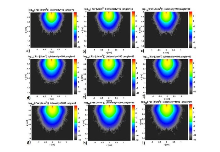
 A wearable wireless tomographic probe has been developed for continuous monitoring of tissue perfusion at different depths. Using the NIRS method, blood ?ow can be continuously measured at different tissue depths. This device has been designed following conclusions of extensive computerised simulations and it has been validated using a vascular phantom.
Berthelot M, Yang GZ, Lo B, 2018, Tomographic probe for perfusion analysis in deep layer tissue, Pages: 86-89, 2018 IEEE 15th International Conference on Wearable and Implantable Body Sensor Networks (BSN).
Article supporters
Article text (excluding photos or graphics) © Imperial College London.
Photos and graphics subject to third party copyright used with permission or © Imperial College London.
Reporter
Erh-Ya (Asa) Tsui
Enterprise