Infection Diagnostics: Miniature Fibre-Optic Probes for Rapid Bacteria Detection

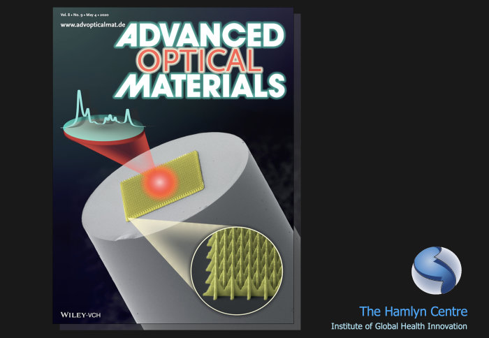
A novel method for infection diagnostics proposed by Hamlyn Centre researchers is featured on the latest Advanced Optical Materials cover.

On 4th May 2020, the latest Advanced Optical Materials cover story featured the novel research conducted by our Hamlyn Centre research team for the applications in infection diagnostics. The team developed miniature fibre-optic probes using a micro-scale 3D-printing technique known as two-photon polymerisation (2PP). Through these probes, live, unlabelled Escherichia coli were detected in solution within a few seconds.
Surface-enhanced Raman Spectroscopy
Raman spectroscopy, widely adopted in various application for identification and characterisation of materials, is a powerful analytical tool for detection of pathogens and biomarkers, and for diagnosis of diseases ranging from infection to cancer in the field of medicine.
However, use of Raman spectroscopy in the clinic has always been challenging since Raman signals are intrinsically weak and can easily be swamped by interfering background signals.
“Surface-enhanced Raman spectroscopy (SERS)" is one of the signal enhancement techniques that can combat this issue and metal surfaces with micro-/nanoscale structures on SERS can offer the SERS effect.
Conventional fabrication methods for SERS effect are well established but developing miniature fibre-optic SERS probes is difficult due to the need for advanced micro-fabrication technologies.
Fibre-Optic SERS Probes Fabricated Using Two-Photon Polymerisation
In this article, our researchers Dr Jang Ah Kim, Dr Dominic Wales, Dr Alex Thompson and Professor Guang-Zhong Yang demonstrated a simple, two-step approach to produce the fibre-optic SERS probes and demonstrated the probes for capability for rapid bacteria detection.
The probes were fabricated by 3D-printing polymeric microstructure on the tips of optical fibres (as thin as roughly 2-3 times the thickness of a single strand of hair) using 2PP technique and then depositing a thin layer of gold.
Using these fibre-optic SERS probes, a significant signal enhancement was measured. Moreover, having optimised the microstructures and characterised the sensing performance of the probes, the team also showed that the probes were able to detect E. coli in solution with short integration times in milliseconds to seconds range.

In the field of medicine, this is the first report of detection of live, unlabelled bacteria using a fibre-optic SERS probe, and indicates the future potential for clinical applications.
The novelty and impact of this work has been acknowledged and thus the article was selected for the front cover picture of the current issue of Advanced Optical Materials (Volume 8, Issue 9, May 4, 2020).
This research was conducted as a part of EPSRC Programme Grant, “Micro-robotics for Surgery (EP/P012779/1)” and was published in Advanced Optical Materials (J. Kim, D. J. Wales, A. J. Thompson, G.-Z. Yang, “Fiber-Optic SERS Probes Fabricated Using Two-Photon Polymerization For Rapid Detection of Bacteria”, Advanced Optical Materials, 8(9), May 2020, 1901934).
Article supporters
Article text (excluding photos or graphics) © Imperial College London.
Photos and graphics subject to third party copyright used with permission or © Imperial College London.
Reporter
Dr Jang Ah Kim
Department of Mechanical Engineering
Dr Alex Thompson
Department of Surgery & Cancer
Erh-Ya (Asa) Tsui
Enterprise