A Smart Surgical Imaging System for Breast Cancer Surgery
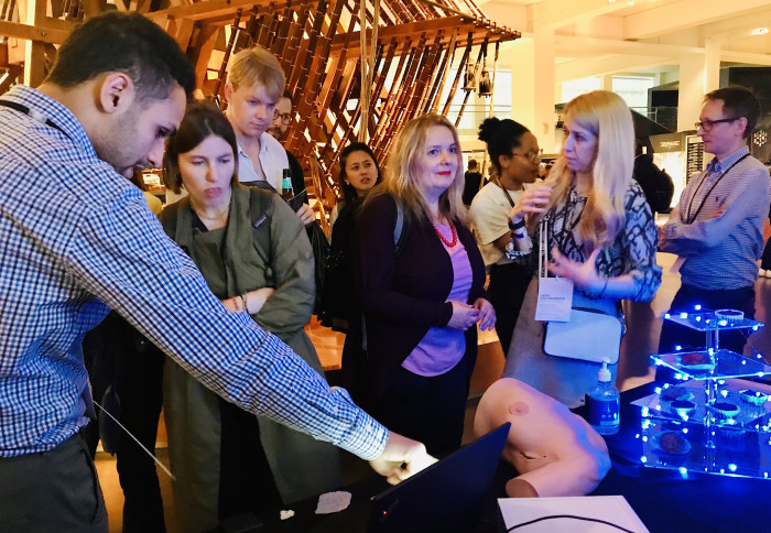
Researchers presented a smart surgical imaging system for breast cancer surgery at the latest Science Museum LATES event.
On Wednesday 25th May evening, right after the opening of the special exhibition "Cancer Revolution: Science, Innovation and Hope
 Led by our clinical reader in breast surgery Daniel Leff, our research team from the Hamlyn Centre and Department of Surgery and Cancer presented a smart surgical imaging system, which could precisely visualise the tumour boundaries and help surgeons resect disease more efficiently, in order to reduce the need for follow-up surgeries.
Positive Margins in Breast Cancer Surgery - A Surgical Challenge
Breast cancer is the most commonly diagnosed cancer worldwide, affecting 1 in 8 women (1 in 7 women in the UK alone) during their lifetime. The majority of breast cancers are treated with breast conserving surgery (BCS).
However, 20% of these surgeries fail to remove all the cancer tissues at the first attempt. This results in the traumatic experience of re-operation for the patients and in the additional annual expenses of £60 millions to the NHS.
To overcome this obstacle, pre-operative planning and intra-operative feedback are both essential. Imaging, optical spectroscopy, bio-impedance or mass spectrometry techniques are currently being clinically trialed in view of addressing positive margins (whereby tumour encroaches upon the rim of tissue surrounding the resection).
Among these techniques, fluorescence guided surgery (FGS) is promising, as it provides real-time intra-operative macroscopic visualisation of the targeted tissue (i.e. tumour), which is easily interpretable.
 "In FGS, a fluorescent contrast agent (fluorophore) is externally administered to the patient, and accumulates in the tumour. Upon the fluorophore’s accumulation, its fluorescence can be captured with an FGS camera system and be utilised to guide tumour resection in real-time" Professor Daniel Elson explained.
A Smart Surgical Imaging System for Breast Cancer Surgery
Our research team from the Hamlyn Centre and Department of Surgery and Cancer developed a smart surgical imaging system for breast cancer surgery by utilising Indocyanine green (ICG) fluorescence image processing techniques, aiming to prevent further re-operations.
Indocyanine green (ICG) is the most widely accepted fluorophore due to its near-infrared (NIR) spectral properties, favourable penetration, depth, and low toxicity.
The lead researcher Dr Maria Leiloglou said: "Our research focuses on the use of an imaging system with two cameras that can acquire colour and fluorescence light emitted from molecules in the cancerous tissue. These images are processed to visualise the cancer boundaries overlaid on the surgical scene."

"Our device was tested in 50 patients with the fluorescent contrast agent (ICG), proving the feasibility of the approach. Currently, we are performing clinical tests with multi-modal optical imaging - combining a new contrast agent ("5 Aminolevulinic Acid") and multicolour information", our clinical research fellow Martha Kedrzycki said.

In this work, our research team also proposed a hybrid model that was trained anew with all the available ground truth data and applied to predict the tissue class (healthy/tumour) on the in-vivo images (tumour in-situ and surgical cavity) within the angiography cohort.
In conclusion, with the long-term vision to improve the future of surgical practice, our research team starts with better breast conserving surgery outcomes. This proposed smart surgical imaging system could potentially guide the surgeons to remove the cancer successfully at the first operation.
"The patients could receive optimal treatment, enabling them to complete their treatment journey, recover and return to work faster. Our technology could be a cost-effective solution for the NHS as it could decrease re-operation costs and optimise the use of staff and infrastructure", Dr Maria Leiloglou emphasised.
(Maria Leiloglou, Martha S. Kedrzycki, Vadzim Chalau, Nicolas Chiarini, Paul T. R. Thiruchelvam, Dimitri J. Hadjiminas, Katy R. Hogben, Faiza Rashid, Rathi Ramakrishnan, Ara W. Darzi, Daniel R. Leff and Daniel Elson, "Indocyanine green fluorescence image processing techniques for breast cancer macroscopic demarcation", Scientific Reports, 12, 8607, May 2022)
Article text (excluding photos or graphics) © Imperial College London.
Photos and graphics subject to third party copyright used with permission or © Imperial College London.
Reporter
Erh-Ya (Asa) Tsui
Enterprise