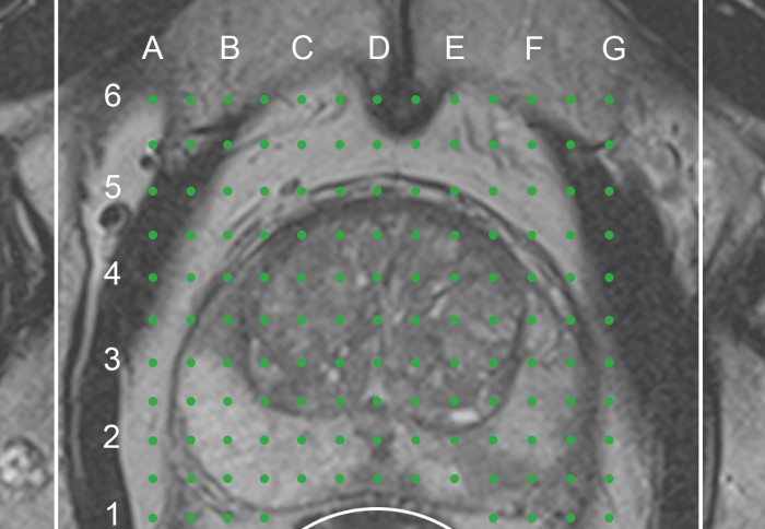
Unique clinical imaging dataset to accelerate diagnosis of prostate cancer

The National Cancer Imaging Translational Accelerator has released a unique clinical imaging dataset from the Prostate MRI Imaging Study (PROMIS).
The NCITA, a national UK infrastructure consortium that brings together nine world-leading medical imaging centres, including one at Imperial College London, in partnership with the ReIMAGINE Consortium, released the imaging dataset, which can be accessed by clinical researchers, initially on an application basis, for artificial intelligence (AI) research to speed up imaging diagnosis of clinically significant prostate cancer using novel machine learning tools.
About the PROMIS study
PROMIS was a landmark multi-centre study, funded by the National Institute for Health and Care Research (NIHR) and led by Professor Hashim Ahmed, and has reshaped the prostate cancer diagnostic pathway. The study assessed the accuracy of an imaging technique called multiparametric magnetic resonance imaging (mp-MRI) for diagnosing prostate cancer compared to a detailed biopsy procedure.
“PROMIS was a once-in-a-lifetime, never to be repeated, validation study in which all patients underwent a transperineal template mapping biopsy of the prostate. This gold standard reference test provides the highest accuracy and fidelity of cancer status and location for a population at risk.” Professor Hashim Ahmed Chair of Urology at Imperial College London
A total of 576 men underwent a mp-MRI scan, followed by a systematic transrectal ultrasound (TRUS)-guided biopsy and a 5 mm transperineal template mapping (TPM) biopsy across the entire prostate. As the MRI scans were reported independently to the biopsy, all participants, underwent a full prostate biopsy, irrespective of the MRI result.
The results published in the Lancet showed that mp-MRI scan was highly accurate in detecting 93% of prostate cancers compared to 43% for the TRUS biopsy test. The mp-MRI scan was also shown to accurately identify about 25% of men who did not have prostate cancer and who might safely avoid having a biopsy.
The imaging dataset can now be accessed by clinical researchers, initially on an application basis, for artificial intelligence (AI) research to speed up imaging diagnosis of clinically significant prostate cancer using novel machine learning tools.
Changes in international guidelines
These landmark results from the PROMIS study and a number of other high-profile studies including PRECISION (PRostate Evaluation for Clinically Important disease, Sampling using Image-guidance Or Not?) published in the New England Journal of Medicine have led to changes in international guidelines for prostate cancer care to reduce the proportion of men having unnecessary biopsies and improve the detection of clinically significant prostate cancer.
The 2019 UK National Institute for Health and Care Excellence (NICE) guidelines and the 2019 European Association of Urology guidelines now recommend that all men with a suspicion of prostate cancer receive a mp-MRI scan as an initial test prior to prostate biopsy. Both organisations also recommend considering avoiding a prostate biopsy in men with low clinical risk of prostate cancer who have a non-suspicious MRI, after an informed discussion with the patient.
Unique clinical imaging dataset for AI and machine learning research
The clinical imaging dataset from the PROMIS study includes data from 11 NHS hospital trusts across the UK, and comprises of over 500 consecutive pre-biopsy mp-MRI scans paired with comprehensive template mapped biopsies of the prostate. This is an entirely unique dataset as it includes paired MRI scanning and template mapped biopsy of the entire prostate, including template biopsy validated negative MRI cases.
The dataset, which has been curated by the ReIMAGINE consortium at UCL, is now accessible to clinical researchers who are developing AI algorithms and machine learning tools for clinical application to speed up prostate cancer diagnosis.
Accessing the PROMIS study dataset
The curated PROMIS study imaging dataset is hosted by NCITA, a UK-wide clinical imaging research infrastructure funded by a 5-year Cancer Research UK Accelerator Award. Professor Eric Aboagye, from the Department of Surgery and Cancer, leads Imperial's involvement in NCITA. NCITA provides a federated digital infrastructure for the secure storage and sharing of imaging data as well as data integration and analysis services using AI and machine learning tools.
The curated imaging dataset is now accessible to clinical researchers on an application basis to The ReIMAGINE PCa Risk Trial Management Group. For further details, please contact by email.
This article was based on a NCITA press release.
Article text (excluding photos or graphics) © Imperial College London.
Photos and graphics subject to third party copyright used with permission or © Imperial College London.
Reporter
Benjie Coleman
Department of Surgery & Cancer