New heart treatment helps the body grow a replacement valve
by Ian Mundell

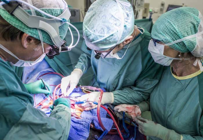
Heart valve replacement is a life-saver, but still has drawbacks (Image: Getty Images)
Replacement heart valves that grow inside the body are a step closer to reality following studies led by researchers at Imperial.
Surgery to replace faulty heart valves has been possible for more than 60 years, but the treatment has medical drawbacks, both with mechanical or biological valves. But what if the body’s natural repair mechanisms could be harnessed to build a living heart valve, right where it is needed? Recent studies led by researchers at Harefield Hospital and Imperial’s National Heart and Lung Institute suggest that this approach is entirely possible.
Heart valve replacement is a life-saving treatment, but it is rarely a long-term solution. Both mechanical and biological valves have their own drawbacks. Patients with mechanical valves must take drugs for the rest of their lives to prevent blood clotting. Biological valves, on the other hand, only last between 10 to 15 years. The treatment is particularly challenging for children with congenital heart defects, as the valves do not grow along with their bodies and must be replaced several times before they reach adulthood.

The new approach developed by Sir Magdi Yacoub’s team at Harefield and Imperial is much more adaptable. “The aim of the concept we’ve developed is to produce a living valve in the body, which would be able to grow with the patient,” says Dr Yuan-Tsan Tseng, a biomaterials scientist working at the National Heart and Lung Institute and the Harefield Heart Science Centre.
The procedure begins with a nanofibrous polymeric valve, but made from a biodegradable polymer scaffold rather than a durable plastic. “Once this is inside the body, the scaffold recruits cells and instructs their development, so that the body works as a bioreactor to grow new tissue,” Dr Tseng explains. “The scaffold gradually degrades and is replace by our body’s own tissues.”
The scaffold material used to make the valve is the key innovation. “It has the capability to attract, house, and instruct appropriate cells from the patient's own body, thereby facilitating tissue generation and maintaining valve function.”
Building up, breaking down
The design and manufacture of the valve is set out in a recent academic paper*, along with validation of its performance in the laboratory and the first results of animal tests. The valves were transplanted into sheep and monitored for up to six months. “The valves performed very well,” says Dr Tseng. “They continued to function for the six months of the trial, and also showed good cellular regeneration.”
In particular, the study shows that the scaffold was able to attract cells from the blood stream which then developed into functional tissues, a process known as endothelial-to-mesenchymal transformation (EndMT). “We’ve also seen nerves and fatty tissues growing in the scaffold, as we might expect in a normal valve.”
Meanwhile, the polymer could be seen degrading to make way for the new tissue. This degradation process was followed with the help of gel permeability chromatography (GPC) in the Agilent Measurement Suite (AMS), a facility in Imperial’s Molecular Sciences Research Hub in White City fitted out with advanced analytical instrumentation provided by Agilent.

“GPC was able to tell us the molecular weight of the polymer in samples taken from the valves at various time points during the in-vivo study,” Dr Tseng says. This showed that the structure was gradually being broken down, but without affecting the valve’s performance.
“If there was no regeneration, the valve would fall apart as the polymer degrades. But what we see is continuing functionality, and that means cell regeneration is taking place over time. That proves that our idea of in-vivo regeneration is working.”
More work is required to determine exactly which processes are causing the polymer to degenerate and how closely it is linked to tissue regeneration. “But the tissue regeneration is definitely sufficient to cover the structural integrity and functionality of the valve,” says Dr Tseng.
On to clinical trials
The next step is to continue the animal studies, to follow the tissue regeneration process for longer. This data will be essential in order to get regulatory approval for the first clinical trials, hopefully in the next five years or so.
Further work will also be required on the processes used to manufacture the valves. “There are various improvements to make on the manufacturing side, and we will potentially use the Agilent Measurement Suite again to help us optimise the polymer, so that it is performing in the desired way,” Dr Tseng says.
The work of Dr Tseng and his colleagues presents an excellent example of the kind of contribution the Agilent Measurement Suite can make. Professor Marina Kuimova Director, Agilent Measurement Suite
This demonstrates the continuing role that the facilities Imperial can play advancing innovation. “At the Agilent Measurement Suite we have a wide range of the state-of-the-art analytical instrumentation and expertise that can serve medical and biological research communities,” says Professor Marina Kuimova, Director of the AMS. “The work of Dr Tseng and his colleagues presents an excellent example of the kind of contribution the Suite can make.”
As work on the replacement valves progresses, the team will also start looking for commercial partners to help them in the later stages of clinical trials. “That requires a different kind of expertise, which we don’t have in academia.”
While the focus at present is replacement heart valves, the approach could have many other applications. “Once you have the scaffold, it becomes a platform technology that you can use to engineer other tissues,” says Dr Tseng. Possibilities include addressing vascular conditions, such as repairing blood vessels damaged in dialysis, and building cardiac patches to repair damage to the heart.
*Yacoub, MH, Tseng, YT, Kluin, J et al. Valvulogenesis of a living, innervated pulmonary root induced by an acellular scaffold. Communications Biology 6, 1017 (2023). https://doi.org/10.1038/s42003-023-05383-z
Article text (excluding photos or graphics) © Imperial College London.
Photos and graphics subject to third party copyright used with permission or © Imperial College London.
Reporter
Ian Mundell
Enterprise