Congratulations to Cristina Lo Celso, winner of the Goeppert-Mayer medal!
by Emily Govan
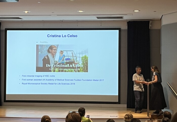
Many congratulations to Professor Cristina Lo Celso on winning the first Goeppert-Mayer medal for intravital microscopy research.
Professor Lo Celso recently attended the Australian Intravital Microscopy Symposium, held in Sydney, where she delivered the keynote lecture. She was awarded the medal at the symposium, for her work using intravital microscopy to uncover new aspects of haematopoietic stem cell biology.
Maria Goeppert-Mayer
Maria Goeppert Mayer was a German-born American theoretical physicist, and Nobel laureate in Physics for proposing the nuclear shell model of the atomic nucleus. She was the second woman to win a Nobel Prize in physics, the first being Marie Curie.
A graduate of the University of Göttingen, Goeppert Mayer wrote her doctoral thesis on the theory of possible two-photon absorption by atoms.
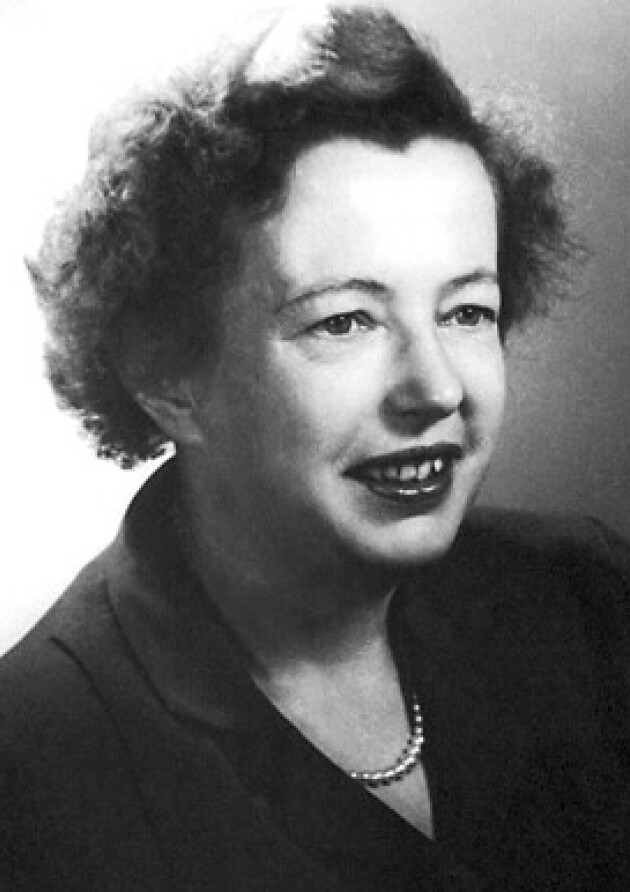
At the time, the chances of experimentally verifying her thesis seemed remote, but the development of the multiphoton laser in the 1960s later permitted this. Today, the unit for the two-photon absorption cross section is named the Goeppert Mayer (GM) unit.
As her PhD was in 1930, it took 30 years for her theory to be put into practice!
This year marks the first award of a medal in her honour given in recognition of work in intravital microscopy research.
Talking to Professor Lo Celso
We caught up with Professor Lo Celso to learn a little about the medal and her work.
Can you please tell us a bit about what you are working on?
“Two photon excitation is the main modality of microscopy that’s used for intravital microscopy, which is looking for cells within live tissues and organisms. The work is even taking place with humans now! I have been using intravital microscopy from 2006 and I am interested in working out how cells live in the bone marrow. Easier to do on mice, using bones on the top of their skull.
This sort of microscopy is far superior to histology as it allows you to see the cells in action. You can learn how much a cell moves, and how it interacts with other cells, for example if they interact for a time or a little bit, for instance in histology you may see a cell close to another one but this position may not be fixed in time. So the two cell types may be close sometimes but then another time they will be further away. Intravital microscopy gives a more dynamic view. This is particularly useful when looking at the bone marrow, where billions of cells are made and sent into circulation every day. It makes sense that dynamic interactions between cells are important to achieve this.
We have found out lots of interesting things, for instance blood stem cells reside in niches, formed by an assortment of other cell types and critical for blood stem cells to work. They are dynamic, they wiggle and project protrusions. They also have a bit of migratory behaviour, they move slowly and takes them a long time, but they do still move across the bone marrow space. One of our unexpected findings is that blood stem cells move a bit faster and bit further when the mouse carries and infection. Blood stem cell migration also explains why, when we look for blood stem cells in sections, they are far apart from each other. We understand through intravital microscopy that this pattern arrives because the cells move from each other.
It is also interesting that leukaemia cells migrate across the bone marrow space. Following chemotherapy, the resistant cells from certain types of leukaemia migrate faster. If we stop the migration then they start dying. Independent groups have initiated clinical trials which affect the movement of these cells. It’s completely different to chemo and is very promising.
We have recently seen that as myeloid leukaemia develops it recruits certain types of immune cells that help the disease to grow. This work initiated with intravital microscopy, leading us to find something unexpected that we are now exploring further to develop potential new, more targeted and less toxic therapies.
"The medal showed me that there is a growing community of people that appreciate all the hard work that is involved in intravital microscopy – it gave me a nice fuzzy feeling!" Professor Cristina Lo Celso
Intravital microscopy is used in many organisms.
One interesting thing I saw at the conference where the medal was awarded, was that people are using the research to study immune cells in the human cornea and finding different cell behaviours in people with hay fever. This new technology is coming out of the laboratory and leading to surprises and discoveries. That was the coolest thing I saw!”
That sounds fantastic. And how did you feel to win this medal?
“This is a newly created medal, and it told me that there is a growing community of people that appreciate all the hard work that is involved in intravital microscopy – it’s nice to see it. The work is challenging and slow-going. We are doing it at a time where the dominant approaches are multi-omics, where you can extract huge datasets and lots of information very quickly. But the work I am doing takes many hours and is very complex and it can take months if not years to generate the images we aim for. To see it recognised gave me a really nice fuzzy warm feeling!”
Article text (excluding photos or graphics) © Imperial College London.
Photos and graphics subject to third party copyright used with permission or © Imperial College London.
Reporter
Emily Govan
Department of Life Sciences