Organ organisation: why sex-based differences matter
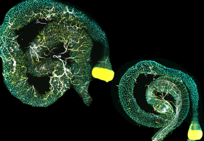
Dissected female (left) and male (right) guts of adult Drosophila. The tracheal branches that normally hold gut loops together are visualise
Organ geometry is not a developmental accident - specific male and female organ configurations are maintained by complex multi-organ communication.
Many diseases manifest differently in males and females, but the molecular and physiological mechanisms underpinning this remain poorly understood.
A new study, published in Nature, has revealed differences in the development, positioning, and ongoing maintenance of organs between male and female fruit flies that have consequences for healthy organ function. Though far removed from human health, the study investigates basic principles of organ development and sex differences.
“The shape and position of organs is not a developmental accident,” said Professor Irene Miguel-Aliaga, a Principal Group Leader at the Francis Crick Institute who initiated the research at the MRC Laboratory of Medical Sciences (LMS) at Imperial College London, and coordinated across both institutions. “They have not ended up where they have ended up inside the body because that’s where they happened to be made.”
Sex-dependent organ organisation
The early development of any animal’s body is an intricately choreographed sequence of molecular interactions that create organisms with great precision. The shape and positioning of an organ amongst its neighbours follow an important logic that goes far beyond a simple jigsaw puzzle of slotting in pieces wherever they fit.
In the latest study, the team centred in particular on the gut, and found active mechanisms maintaining gut shape and position. Professor Miguel-Aliaga said: “We have found that there is a complex dialogue between the gut and its vessels that actively keeps the gut looped in adult animals and also makes gut shape different between the sexes.
“Somewhat unexpectedly, the main role of these vessels (so-called trachea in flies) does not seem to be in delivering oxygen to the intestine (what they are known for in other contexts) but rather in literally holding, or ‘stitching’ gut loops together.”

The researchers explain that the maintenance of this position may have broad health implications other than simple function. “Gut shape may matter to flies,” says Dr Laura Blackie, one of the two lead authors of the study. “Using genetic tricks, we can relax the shape of the gut of the fly, or even make female flies with male gut shape regulators. These flies have reduced fecundity, and we now want to understand why.
“Because this gut relaxation does not seem to affect the intestine itself, we have an idea that by changing gut shape and/or position we may be interfering with the messages the intestine exchanges with other organs, and that this is what might impact fecundity. But we now need to test this.”
The study suggests that the positioning and interaction between different organs influences overall health in ways specific to each sex, and at the whole-body level rather than being restricted to each isolated organ.
The findings also showed sex differences in parts of the body completely separate to the reproductive organs.
Dr Pedro Gaspar, co-lead author of the study, said: “Signals from the gut muscle influence the development of sex-specific vascular structures, which in turn shape the gut. This suggests that sex differences in body structure can arise not just from traditional sex hormones produced by the gonads but also from signals and mechanical effects originating from other body tissues.”
According to the researchers, this development of the technology could provide new opportunities to investigate inter-organ communication.
Dr Gaspar added: “We have advanced the idea that inter-organ physical tethering is critical to maintaining organ shape and is modulated by sex-determining factors. We have unlocked a new set of tools in 3D imaging that will enable the future modelling of signals involved in inter-organ communication and the impact of organ adjacencies in metabolism and physiology."

A flying start
The biology and bio-geometry of flies may be far removed from human health, but the researchers highlight there is a reason this versatile model species is so valuable.
Professor Miguel-Aliaga explained: “It is exciting to me that we can assess the impact of very precise, spatiotemporally controlled genetic interrogations at the level of a whole-body multi-organ network – I don’t think we could have done this in any other experimental system at the scale at which we have done it in flies.”
The study involved a series of cutting-edge techniques and a team effort calling on broad specialty skills, reaching from 3D scanning to genetic engineering and mathematical modelling.
“We first had to acquire 3D scans of many (thousands!) of flies,” said Professor Miguel-Aliaga. “Then we developed ways to quantify organ shape, position and adjacencies between organ, in collaboration with the Mahadevan lab at Harvard. Finally, we combined these scans and quantitative approaches with sophisticated genetic manipulations that allowed us to determine what organs are talking to each other, and what molecular and mechanical messages they exchange.”
From flies to humans
You may expect that something as fundamental as differences between male and female organ positions would be well understood. But, the team say, examining the details of the positioning and molecular interactions required techniques only now available.
“We had to be able to scale up both the imaging and its quantification so that we could look at variation: what is stereotypical across individuals, and what is shared or different between the sexes,” said Professor Miguel-Aliaga. “Our tools to genetically interrogate complex systems have also become a lot more granular and sophisticated, which helped with our mechanistic explorations.”
The researchers explain that potential next steps of the research could be to explore the mechanisms and principles of sex differences in humans, which may ultimately help to better understand disease and help to improve treatments.
“We are beginning to apply the methods we have developed in flies to human MRI scans. We can now quantify 3D intestinal features of our own intestinal tract. We do not know much about possible sex differences in our gastrointestinal tract, despite the fact that many gastrointestinal disorders are sex-biased in their incidence and/or severity. Perhaps by applying our quantifications of 3D features to our own intestine in its intact three-dimensional arrangement we may be able to find some features that are predictive or diagnostic of disease.”
Read the full study in Nature: https://doi.org/10.1038/s41586-024-07463-4
Article supporters
Article text (excluding photos or graphics) © Imperial College London.
Photos and graphics subject to third party copyright used with permission or © Imperial College London.
Reporter
Lindsay Keith
Institute of Clinical Sciences