Visualizing molecular engineering

Imperial researchers share their best images featuring the field of molecular science and engineering for IMSE's image competition
Earlier this year, the Institute for Molecular Science and Engineering (IMSE) asked researchers at Imperial College London, from undergraduates to staff, to share their favourite scientific images for a competition.
The images had to be original and showcase the transdisciplinary field of molecular science and engineering. Among the entries, we received portraits of researchers doing experiments in the lab, AI generated visions of molecules, images of cells taken with a fluorescence microscope, high resolution pictures from a scanning electron microscope (SEM), magnified images of batteries’ surface and photos of patterns appearing on polymers under varying conditions taken with an optical camera!
The IMSE Operations Team cast their vote anonymously and chose 3 winners who received both a monetary prize and a printed copy of their image during IMSE’s 2024 Research Showcase.
Zain Ahmed, from the Polymers and Microfluidics research group, won the competition with the image: “Look closer into the rainbow amphitheater”. On his own words, Zain explained that when a rubber-like polymer is exposed to plasma, spontaneous patterns form on its smooth surface. In a trapezoidal sample, these patterns align in a manner resembling rows in an amphitheatre. When illuminated from above with white light, vibrant rainbow colours appear due to the diffraction caused by the surface patterns.
Dr Cinzia Klemm, from the Bioengineering Department, won the second prize with her image showcasing bioproduction. With the title “Vitamin Y(east) – Sustainable production of beta-carotene in S. cerevisiae”, Cinzia’s image shows Saccharomyces cerevisiae cells engineered to produce beta-carotene, a vitamin A precursor. Beta-carotene is auto fluorescent (shown in yellow) and accumulates in the lipid bodies of the cell. Due to its autofluorescence, beta-carotene is an excellent high-value product for studying bioproduction in single cells. Cell walls are stained using calcofluor white (cyan).
Elman Huseymov, MRes in Green Chemistry, was joint-third with his image entitled “Tris(acetylacetonato) iron (III)-(Fe(acac)3) in Dimethylbutylammonium hydrogen sulfate - ([DMBA]+[HSO4]-)”. Elman took this image when he was investigating the solubility of different iron sources in an ionic liquid dimethylbutylammonium hydrogen sulfate ([DMBA][HSO4]). The image demonstrates that Fe(acac)3 did not dissolve.

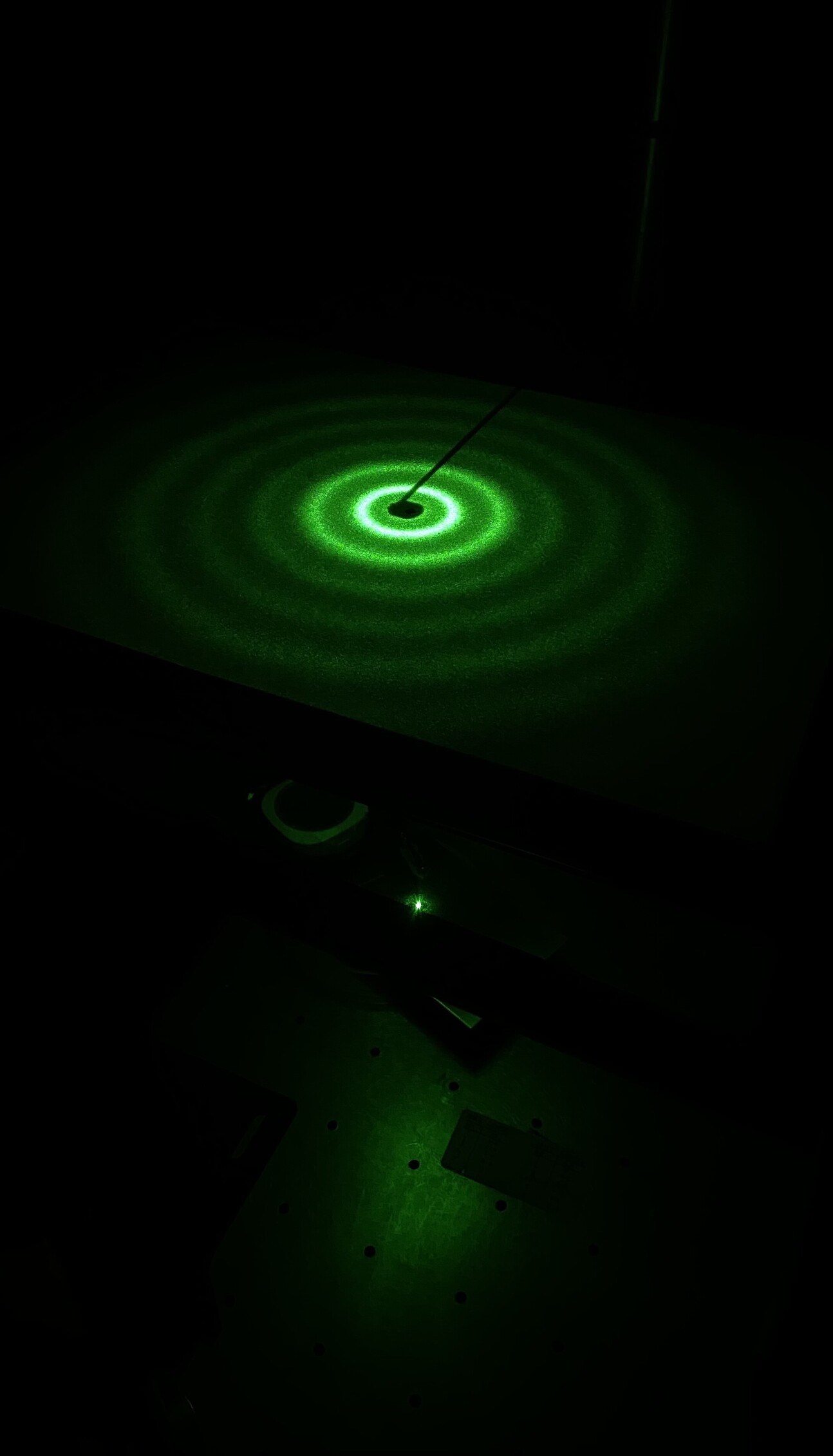
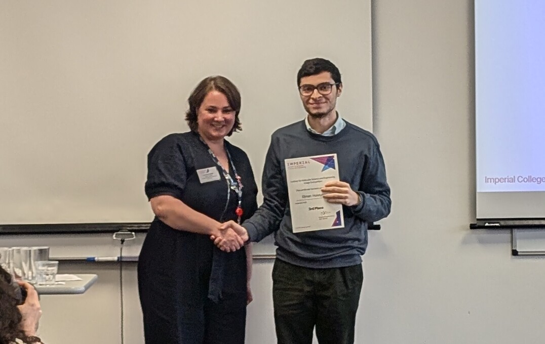
Zain also won joint third with another image, entitled “Wrinkled airy disk”, an additional example of patterns appearing on polymers when the conditions change. “A thin, standard polymer develops labyrinth-like surface patterns when subjected to plasma. When a green laser is directed onto the surface, the light diffracts, and the resulting diffraction pattern is projected onto a screen as concentric rings” Zain explained to the audience at the Research Showcase.
We hope you feel inspired by these beautiful images of research and will be ready to snap a photograph next time you are in awe of nature and science!
Thank you to all participants for sharing their beautiful images of research with IMSE.
Article text (excluding photos or graphics) © Imperial College London.
Photos and graphics subject to third party copyright used with permission or © Imperial College London.
Reporter
Elena Corujo Simon
Faculty of Engineering


![Upon investigation of the solubility of different iron salts in an ionic liquid, namely [DMBA] [HSO4], the image demonstrates that Fe(acac)3 did not dissolve.](/newsarchive/images/mainnews2012/e-huseynov-1_1727772735267_x2.jpg?r=3875)
