New MRI scanner to help better detect knee injuries

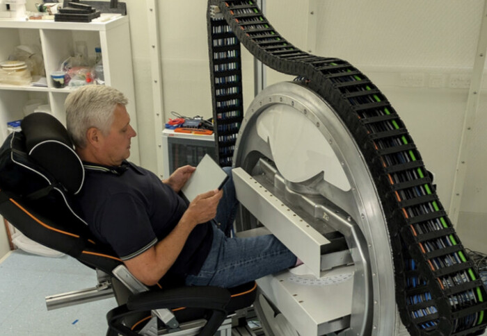
Imperial researchers have developed a novel low-cost MRI scanner that exploits the "magic angle effect" of MRI scans to better detect knee injuries.
"I am very proud that a collaboration between Imperial's different departments has led to this development and anticipate it will help the diagnosis of partial anterior cruciate ligament tears." Mr Chinmay Gupte Clinical Reader, Department of Surgery and Cancer
A collaboration between radiology engineers in Imperial's Department of Mechanical Engineering, surgeons in the Department of Surgery and Cancer, and radiologists from the NIHR Imperial Biomedical Research Centre, has led to a potentially groundbreaking study on the first in vivo images from magic angle directional MRI imaging for anterior cruciate ligament injuries of the knee. The study was published in the journal Magnetic Resonance in Medicine.
Researchers have developed a novel low-cost MRI scanner that exploits a physical phenomenon known as the "magic angle effect" of MRI scans. This used to be considered as a nuisance effect, causing distortions of signal in conventional MRI. However, led by Mr Chinmay Gupte, Dr. Mihailo Ristic and Dr Dimitri Amiras, the team consisting of Karyn Chapell, Harry Lanz and John McGinley, has managed to apply the physics of this effect to image individual collagen fibres of the anterior cruciate ligament and meniscal cartilages of the knee.
It is anticipated that this will help diagnosis of partial anterior cruciate ligament tears in sportsmen and women. As the MRI scanner is low field and occupies very little space, the installation and operational costs of the scanner are a fraction of the conventional MRI scanner. This may also reduce the cost of MRI scanning in the NHS.
Speaking about the research, Mr Chinmay Gupte, a consultant orthopaedic sports knee surgeon and clinical reader at Imperial, said: "I am very proud that a collaboration between Imperial's different departments has led to this development and anticipate it will help the diagnosis of partial anterior cruciate ligament tears.
"We are now beginning larger scale clinical trials to look at the individual collagen fibres in the anterior cruciate ligament and medial cartilages of the knee to better understand injuries to the structures and improve outcomes in the hundreds of thousands of people who suffer these injuries every year."
The study continues and is now part of the Medtech acceleration scheme at Imperial.
First in-vivo magic angle directional imaging using dedicated low-field MRI. Mihailo Ristic, Karyn E. Chappell, Harry Lanz, John McGinley, Chinmay Gupte, Dimitris Amiras. Magnetic Resonance in Medicine. 20 October 2024. https://doi.org/10.1002/mrm.30332
Article supporters
Article text (excluding photos or graphics) © Imperial College London.
Photos and graphics subject to third party copyright used with permission or © Imperial College London.
Reporter
Benjie Coleman
Department of Surgery & Cancer