New AI stroke brain scan readings are twice as accurate as current method
by Samantha Rey

AI pinpoints stroke timing, treatment potential from a single scan
New AI software can read the brain scans of patients who have had a stroke, to more accurately pinpoint when it happened and help doctors work out whether it can be successfully treated.
It is hoped that the new technology will ultimately enable faster and more accurate emergency treatment of patients in a hospital setting. Knowing the time the stroke started is important because standard treatments only work in the very early stages post-stroke – and may otherwise cause secondary damage.
The software, devised by researchers from Imperial College London, Technical University of Munich, and Edinburgh University, addresses two of the most difficult challenges in assessing stroke patients – identifying the onset time of the stroke and whether the damage can be reversed. The software has been found to be twice as accurate as the current method – a visual assessment of the scan by a medical professional, who considers how dark a stroke area appears on CT scans of the brain.
A stroke occurs when the blood supply to part of the brain is blocked or reduced, preventing brain tissue from getting oxygen and nutrients. Brain cells then start to die quickly.
As time progresses, some treatments become ineffective or may even cause more problems. Yet finding out when the stroke happened is very difficult. Some strokes may start while the patient is asleep and some patients may have difficulties communicating because of the stroke symptoms.
Dr Paul Bentley at Imperial’s Department of Brain Sciences, who led the research study, said: “For the majority of strokes caused by a blood clot, if a patient is within 4.5 hours of the stroke happening, he or she is eligible for both medical and surgical treatments. Up to six-hours, the patient is also eligible for a surgical treatment, but after this time point, deciding whether these treatments might be beneficial becomes tricky, as more cases become irreversible. So it’s essential for doctors to know both the initial onset time, as well as whether a stroke could be reversed.”
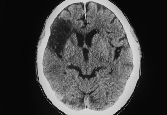
Patients who arrive at hospital with a suspected stroke immediately undergo a CT brain scan, which doctors review to assess how dark the affected areas, or lesions, in the brain are. Darker lesions mean the stroke has progressed further. From this they make an estimate of the time the stroke happened in the past, and whether it may be reversible, and use this to make treatment decisions.
But because all brains are unique, it is very hard to predict with accuracy when the stroke started. Even if doctors know an approximate chronological start time, an individual’s blood flow or blood vessel structure may mean the stroke is progressing more quickly or slowly than average.
The AI algorithm was developed in partnership with Professor Daniel Rueckert (Imperial College London and Technical University of Munich) and Dr Grant Mair of Edinburgh University, who provided expert brain scan interpretation for the algorithm's training phase.
The model was trained on a dataset of 800 brain scans where the stroke time was known. As well as automatically extracting the relevant area from the brain scan, the algorithm reads and analyses the identified lesions, producing a time estimate.
When it was tested on almost 2000 different patients, researchers found the AI software was twice as accurate as using a standard visual method. They believe this is because it includes additional features from the scans, such as texture, and accounts for variations within the lesion and background. It wasn’t only good at estimating the chronological time of the stroke, but also the biological age of the lesions, by which is meant whether they may be reversible.
Dr Bentley, who is also a consultant neurologist at Imperial College Healthcare NHS Trust, which has one of eight hyper acute stroke units in London, explained: “Having this information at their fingertips will help doctors to make emergency decisions about what treatments should be undertaken in stroke patients. Not only is our software twice as accurate at time-reading as current best practice, but it can be fully automated once a stroke becomes visible on a scan.”
Lead author Dr Adam Marcus said: “We estimate that up to 50% more stroke patients could be treated appropriately with treatments because of our method. We aim to deploy our software in the NHS, possibly by integrating with existing AI-analytic software that is already in use in hospital Trusts.”
The research study was funded by NIHR for the purpose of NHS benefit, as well as by Imperial’s Centre for Doctoral Training for AI in Healthcare, and the Graham-Dixon Charitable Trust. The researchers were supported by the NIHR Imperial Biomedical Research Centre, a translational research partnership between Imperial College Healthcare NHS Trust and Imperial College London, which was awarded £95m in 2022 to continue developing new experimental treatments and diagnostics for patients.
Deep learning biomarker of chronometric and biological ischemic stroke lesion age from unenhanced CT is published in NPJ Digital Medicine, DOI 10.1038/s41746-024-01325-z. https://www.nature.com/articles/s41746-024-01325-z
Article supporters
Article text (excluding photos or graphics) © Imperial College London.
Photos and graphics subject to third party copyright used with permission or © Imperial College London.
Reporter
Samantha Rey
Communications Division